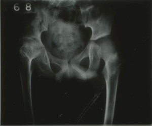
The radiographs from the Stannington collection allow for a detailed insight into the effects that tuberculosis had on the body. One such example is patient 132/1951, a nine year old individual transferred from Fleming Memorial Hospital to the Stannington Sanatorium with Tuberculosis of the Hip. There are a total of 31 radiographic images allotted to this individual, mostly of the pelvic area but also some of the right knee.

Infection is evident in the right hip, the femoral head (head of the femur) and the acetabulum (socket of the pelvis) show visible signs of bone destruction which continues down into the lower part of the pelvis, the ischium. The reduced gap in the joint between the femoral head and the acetabulum is also indicative of tuberculosis (Figure 1). There is also some porosity shown in the femoral head, which displays the weakened state of the bone due to the extent of the infection.
This can be compared to the healthy, left side of the pelvis where a clear ball shaped femoral head can be distinguished with structured shape, lined up with the acetabulum. The gap between the upper and lower sections of the pelvis is evidence of this being a child as the pelvis has not yet fused.
Due to the level of destruction to the right hip, there would have be a significant impact on this individual’s standard of living, the possibility of reduced mobility due to ankylosis of the hip and atrophy.

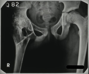
Figure 2, however, shows the results of a surgical intervention to repair damage caused by the tuberculosis infection, a procedure known as arthrodesis. This procedure is descried in the patient’s notes in a letter from the surgeon at the Royal Victoria Infirmary, Newcastle to a doctor at Stannington:
‘… a cortical graft was taken from the anterior aspect of the right tibia. This would was closed with catgut and silk-worm gut to the skin. The right hip was approached from an incision over the posterior aspect of the greater trochanter…. The shaft of the femur was divided just below the greater trochanter and a gap made in the ischium. The bone graft was inserted into two gaps between the femur and the ischium…..’
As a result of the surgery it is noted that there was some flexion in the right hip and some apparent lengthening of the right leg. The tuberculosis infection was deemed quiescent and this individual, after being monitored as an outpatient, went on to be discharged as ‘healed’.
View more radiographs on our Flickr stream https://www.flickr.com/photos/99322319@N07/sets/72157648833066476/
