Stannington’s Reach

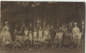
Although based in Northumberland, Stannington Sanatorium wasn’t restricted to taking patients solely from the County of Northumberland.  Looking through the patient files and the earlier minutes of the sanatorium we see that there were many different local authorities wishing to send children to Stannington.  Over the years the authorities of Cumberland, Durham, Newcastle, Gateshead, Rochdale and West Yorkshire all sent patients there at some point, reflecting the uniqueness of Stannington, particularly in its early days, as a sanatorium that catered for children only.  Local authorities would pay for so many beds, and often on the discharge of one child would immediately send another in their place.
 

HOSP/STAN/11/1/57
HOSP/STAN/11/1/57


Opening in 1907 Stannington began life at a time when changes were beginning to be seen in healthcare provision nationally.  Only a few years later in 1911 the National Insurance Act came into force allowing the employed to benefit from medical care on a contributory basis, with particular note made to the treatment of tuberculosis.  We see very few private patients in Stannington throughout its whole history and the majority of children would have been sent by their local authorities as part of the poor relief system, later called public assistance, up until the introduction of the NHS.  Without the assistance of the local authorities many of these children would not have received any medical help at all, and their reliance on them is seen in 1916 when one girl suffering from tubercular patches on her face comes to the end of the time that has initially been paid for by Newcastle Corporation but medical staff consider it appropriate for her to continue to stay on at the sanatorium as her treatment remains incomplete.  However, despite an application being made for an extension Newcastle Corporation refuse to pay and instead the matron makes pleas to the sanatorium’s management committee to allow the girl to stay free of charge until she is fully recovered on the basis that she is a good worker.

“She is a capital worker & is quite healthy in all other ways but her face.  I was wondering gentlemen if you would give permission for this girl to stay on here for some time for free – she could work for us in return for the treatment.” Matron


Given the limited resources of both the sanatorium and the local authorities and considering how rife tuberculosis was during this period it seems quite fair to assume that the children that were eventually admitted to Stannington were the lucky ones, with many more not being able to go.

Differential Diagnosis: Perthes’ Disease

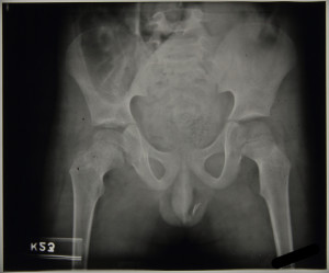
A number of the patients admitted to Stannington were mistakenly given an initial diagnosis of tuberculosis or were found upon examination to be non-tuberculous and were instead allocated an alternative diagnosis. Perthes’ Disease was the most common differential diagnosis assigned to the bones and joints in Stannington, affecting the hip joint this condition was often mistaken as tuberculous-arthritis of the hip.

Perthes’ Disease, a condition that usually affects children between the ages of 4 and 10, is a condition where the blood supply to the femoral head is temporarily lost. This causes the bone at the epiphysis of the femur to soften and breakdown, known as necrosis, giving the femoral head a flattened appearance.

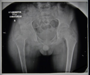
Patient 88/38, a 6 year old boy, is one example of a patient from Stannington with Perthes’ Disease, in this case affecting the left hip. Admitted to Stannington on 31st October 1941, clinical notes read:

‘L. hip – some wasting thigh muscles. Some limitation flexion. Hip in good position.’

FIGURE 2: HOSP-STAN-07-01-02-569_03
Figure 1: HOSP-STAN-07-01-02-569_02


This is supported by the radiographs for the patient, figures 1 and 2 respective of date, and x-ray report:

‘13/11/1941 – Hip – flattened epiphysis has progressed since last x-ray 15/09/1941. Perthes’ Disease

20/11/1941 -Marked flattening of epiphysis L. hip. Some softening of neck. Definite Perthes’ Disease.’


By comparison, tuberculosis of the hip (see posting from 08/12/2014) results in the gradual destruction of the hip joint beginning with a reduction in the joint space between the femoral head and the acetabulum, leading to possible porosity and eburnation in the affected bones and the possibility of pathological dislocation, deformity and loss of use in the affected joint. Even after the disease has reached quiescence it is possible that the individual will suffer with ongoing osteoarthritis or ankyloses.

FIGURE 3: HOSP-STAN-07-01-02-591_03

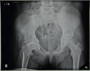
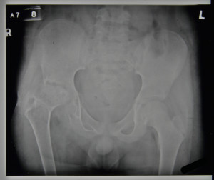
Figure 2, above, is of patient 88/38 with Perthes’ Disease, which can be compared with figure 3, patient 89/21, an individual with TB of the right hip. From the images, note the difference between the Perthes’ Disease where the femoral head becomes flattened and the epiphysis appears to pull away from the metaphysis but generally keeps its ‘ball and socket’ joint appearance with the pelvis compared to the loss of definition of a clear joint with the tuberculous hip, which shows loss of joint space and rarefaction.


For further radiographic images check out the Radiographs from Stannington Flickr Stream athttps://www.flickr.com/photos/99322319@N07/sets/72157648833066476/


Sources

The Perthes’ Association (2011). ‘What is Perthes’ Disease?’ http://www.perthes.org.uk/what-is-perthes-disease/

Surgical Procedures – Osteotomy

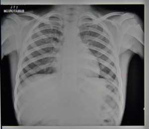
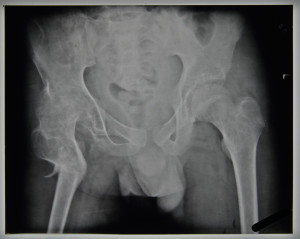
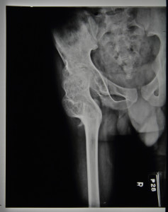
Patient 91/10 was a 15 year old boy admitted to Stannington on 31st October 1941, a letter attached to his discharge report alludes to him having been transferred to Stannington from Newcastle. His initial examination notes reveal he had some tightness over the abdominal area and the right hip was in a state of 45° flexion with some adduction, slight exterior rotation and wasted muscle. Signs of two previous sinuses were also present on the right thigh. The patient had previously been admitted to Stannington from August 1936 until February 1939 when he was discharged in a splint which he wore until the age of 13 prior to his second admission and his hip became flexed; unfortunately we do not hold records for patients this early on and as such have no further details about his previous stay. Figures 1 and 2 are two of the radiographs of his first stay in Stannington, the first showing some tuberculous activity in the lungs and the second showing tuberculous involvement in the right hip joint.

FIGURE 1: HOSP-STAN-07-01-02-686_12
FIGURE 2: HOSP-STAN-07-01-02-686_10


This boy was diagnosed with 3rd stage TB of the right hip and was put forward for a surgical procedure known as an osteotomy. An osteotomy, known as ‘bone cutting’, is a procedure whereby the bone near a damaged joint is cut to realign load bearing surfaces. In the case of patient 91/10 a right femoral osteotomy was performed on the proximal femur to better align it with the acetabulum (socket) of the pelvis, most likely to improve functionality of the limb.

Initial notes on this patient from early 1942 are vague referring to the levels of flexion and adduction in the hip joint but little else. The x-ray report prior to the osteotomy note that no infection is active in the lungs and that there was firm ankyloses in the hip. The osteotomy took place on 13th February 1942.


FIGURE 3: HOSP-STAN-07-01-02-686_05
FIGURE 4: HOSP-STAN-07-01-02-686_04


Figures 3 and 4, respectively, show the patient’s condition before and after the osteotomy was carried out. Figure 3 shows tuberculous infection in the hip joint, affecting the femoral head with some acetabulum involvement. The right hip appears on a slant in comparison to the left sitting higher up, this could potentially have caused the patient to have limp. Figure 4, taken on 14th April 1942, shows the results of the osteotomy, where the femoral head has been repositioned with the aim of realigning the hip joint.

Following the procedure, recovery appears to have gone relatively smoothly. A plaster splint was applied to the patient on 3rd March 1942 where it was noted that the femur had ‘40° abduction – no change’.

No complications are outlined within the notes and the x-ray reports state that there was:

                        ‘14/4/42 –            Good bony union after osteotomy. Position satisfactory.

                        17/7/42 –            Chest no change.

                                                   Hip – osteotomy satisfactory good ankylosis.’


By June 1942 the patient was capable of standing and putting weight on the right leg and is noted to be walking well by mid-July, meriting the osteotomy a great success. The patient was discharged on the 24th July 1942 as quiescent after a 38 week stay at Stannington. The final surgical outcome can be seen in Figure 5.

FIGURE 5: HOSP-STAN-07-01-02-686_14


To see more radiographic images from Stannington, have a look at our Flickr Stream https://www.flickr.com/photos/99322319@N07/sets/72157648833066476

Sources:

Tuli, S.M (2004) Tuberculosis of the Skeletal System: Bones, Joints, Spine and Bursal Sheaths. Jaypee Brothers Publishers.