🎄Christmas Opening Hours for 2024🎄
CLOSED between 4pm Friday 20th December 2024 and 10am Thusday January 2nd 2025
Ordinary opening hours apply before and after this period.

Genitourinary TB – Part 1

Genitourinary TB is the most common form of extra-pulmonary TB today, although the proportion of children in Stannington suffering from this form of TB is relatively low.  Symptoms can include fever, increased urination, and blood in the urine.  In children it is most commonly found either amongst young infants or not until a child reaches puberty and is also a leading cause of congenital TB in new-born babies.


Patient 116/1947 was a 13 year old boy, admitted to the sanatorium on 23 September 1947, and diagnosed with genitourinary tuberculosis.  He had been suffering from a range of medical problems for the past three years, having had a perinephric and a subnephric abscess in December 1944, which was treated with penicillin, and in July 1945 he had a right nephrectomy where the kidney that was removed was found to be tuberculous.  Three months later in October 1945 he returned to the hospital with a right sided epididymitis and again in January 1946 reporting a history of a right sided scrotal abscess which had discharged and healed leaving some thickening at which point haematuria was noted.  He was admitted to hospital again in September 1946 in connection with the right sided epididymitis.


As early as February 1946 it was recommended that he be admitted to a sanatorium and correspondence between the local authorities and Stannington Sanatorium shows that the Administrative Officer of Cumberland County Council was persistent in his attempts to have the boy admitted only to be told by the sanatorium’s Medical Superintendent that there were currently no beds and they were waiting for a suitable side ward to accommodate him.  On his eventual admission he complained of a dull aching pain on the left side of his abdomen, had recently complained of pain on micturition (urination), and was also urinating very frequently, particularly at night.  There was no blood or albumen in the urine at this point, no tenderness felt on the left side of the abdomen, and a small hard nodule about the size of pea was seen in the left epididymis.  His general condition throughout his stay was deemed to be good and chest x-rays were clear of any signs of tuberculosis.


It was decided that given his strong symptoms further investigations of the renal tract were necessary for which he would have to be sent to the RVI in Newcastle as the Sanatorium did not have the required facilities.  Described in his notes as “a perfect nuisance on the ward”, it was decided that he should be sent home to wait for a bed at the RVI.  He was discharged on 19 December 1947.

WWII And The Move To Hexham Hydro

Yesterday the Stannington Sanatorium Project team took a trip to Hexham Hydro, now the Queen Elizabeth High School, to have a look around the building and the grounds as the children from Stannington Sanatorium were moved down to Hexham during WWII as it was deemed to be safer.  After visiting it is easy to see why the Hydro building was chosen by the Sanatorium Committee as it is in beautiful surroundings with views over to Hexham Abbey and the large open rooms making it ideal for the sanatorium’s needs.  The building also has its own walled garden, still well looked after and in use today by the school’s students, with evidence that the sanatorium patients grew produce there which they then went on to sell to local businesses in Hexham.  The Hydro building began life as a private house built in 1859 and known as Westfield House, but was later purchased by the Tynedale Hydropathic Establishment Company and alterations were made so that it could open in 1879 as the Tynedale Hydropathic Hotel.  Over the years additions were made including the large glass Winter Gardens, which would have been used by the sanatorium patients, and many famous clientele reportedly visited including Charlie Chaplin and Ramsay MacDonald. The Hotel eventually went into decline allowing it be used by the sanatorium during the war years as well as acting as a army billet and services bakery.


Read more below to see how WWII affected Stannington:


WWII broke out on 1 September 1939 with the UK officially entering the war 2 days later on 3 September.  Comments made by the matron in the annual report for 1939 highlight the immediate affects the war had on the Sanatorium:

 “…So rapid has been the growth of the Sanatorium that almost every year there has been some change in the structure or equipment to report, but all the changes have been for the securing of that first high ideal – the stamping out of tuberculosis in children.

Now war has come and much has changed.  At any moment a great strain may be put upon our hospitals, and we have had to open wide our doors and be ready to receive 218 adult patients in addition to our own 311 children.  We already have over 100 adult patients in residence, and among them are a number of men of the forces who either from accident or sickness require medical attention.” [HOSP/STAN/1/3/6]

The encroachment on space that the sanatorium had taken for granted for so many years was felt by all.  In the same year the school was evicted from its buildings to make way for beds and lessons were initially undertaken outside on the verandahs until more suitable accommodation was found in the small side wards.


Like people across the country the staff and patients contributed to the war efforts despite the illness faced by the children and additional pressures on the staff.  In 1940 the schoolchildren knitted over 100 woollen comforts for soldiers and 3 large blankets and together staff, children, and friends of the sanatorium raised £352, 17, 1 for the War Savings Scheme as well as additional monies for the Finland Fund, Lord Mayor’s Air-Raid Distress Fund, and the Greek Relief Fund.


After managing to continue operations for nearly two years at Stannington it was decided in 1941 that it was necessary to evacuate the children to a safer place.  The Hydro at Hexham was eventually settled on and over 200 children were moved on 11 August.  The Hydro lacked the vernadahs that were so common in Stannington for open-air treatment but was seen to be a suitable location owing to its lofty rooms, large windows, and beautiful surroundings.  The capacity at Hexham was significantly less than the facilities at Stannington and so the number of patients treated during the war years declined.


It was not until January 1945 that patients and staff were able to return to Stannington on a permanent basis.  Whilst early reports of the sanatorium’s time at Hexham appeared positive it is clear that by 1944 and continuing into the post-war years, the stress of the move and in particular shortages of nursing and domestic staff took its toll on the whole operation.  The 1944 annual report describes how that due to this the full operation of the sanatorium was prevented and consequently the number of patients treated was reduced further still following the initial curtailment felt following the move to Hexham.

Patient 81/39 – A Questionable Diagnosis?

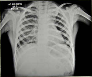
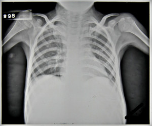
Patient 81/39, a five year old boy, was admitted to Stannington in December 1937 due to ailing health following a two month period in bed suffering from mumps. He had developed a cough, was easily tired and was losing weight. The initial x-ray reports detail a blocked apex in the left lung and marked mottling in the right lung leading to an initial diagnosis of Pulmonary TB, Figure 1. However, following his admission further symptoms started to manifest themselves which indicated that the diagnosis of this patient was more complex than it was initially considered to be.

HOSP-STAN-07-01-02-91_01
FIGURE 2: HOSP-STAN-07-01-02-91_01
HOSP-STAN-07-01-02-91_15
FIGURE 1: HOSP-STAN-07-01-02-91_15


HOSP-STAN-07-01-02-91_14
FIGURE 3: HOSP-STAN-07-01-02-91_14


In April 1938, it was noted the patient had two subcutaneous abscesses on the iliac crest and the knee. A sample of the mucus taken from the abscess on the hip was sent for bacteriological examination. Results of this testing were as follows:

scanty pus cells and much granular debris. No definite organisms seen and tubercle bacilli not found.’

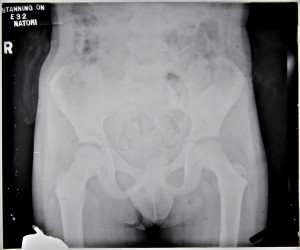
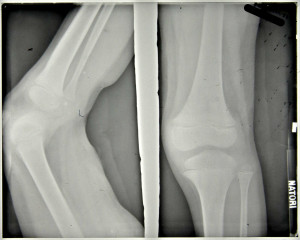
Furthermore, periostitis was noted in the upper end of the ulna which ‘appears septic’ but was regarded as being non-tuberculous. The patient still suffered with a cough but sputum tests were negative and notes state that no tuberculosis was seen. At this stage the x-ray report indicates that no bone lesions are seen in either the leg or the iliac crest, Figures 2 and 3.

Throughout the rest of 1938, the patient’s condition is very variable. An additional abscess is noted in the lumbar region with slight discharge and the apex of the left lung becomes more blocked with the lower lobe of the right lung being described as having been ‘studded with deposits’, however, the sinuses in the thigh and gluteus region are healed.

HOSP-STAN-07-01-02-91_19
FIGURE 4: HOSP-STAN-07-01-02-91_19
FIGURE 5: HOSP-STAN-07-01-02-91_13


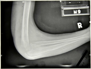
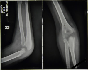
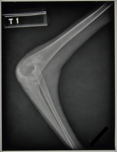
The main focus of the notes centre upon the right elbow which, in September 1938, was described as being very active with discharging abscesses; periostitis was greatly increased in the ulna and also present in the humerus with the joint being ‘badly involved’, see Figure 4. In November 1938 large sequestrum was removed from the elbow, at this time all lesions were considered very active. The elbow continued to be active with an increasing number of ulcers noted to have appeared; a maximum of four seen in February 1939 including one in the right cubital fossa which is incised to produce ‘copious…pus’, Figure 5.

X-ray reports from September 1939 read as follows:

11/9/39 –              Ulna hollowed out to cavity

                            Radius dislocated upward & forward

                            Lower end humerus eroded & partly destroyed.

15/9/39 –            Ulna – upper end partially destroyed, disorganisation of elbow joint’

No further comment is made regarding a diagnosis of tuberculosis in the elbow.


In addition to ongoing changes in the elbow an abscess appeared on the right mastoid, which was opened and drained in October 1938 and is noted to have become less active by November 1938. However, this abscess continued to open throughout the patient’s stay at Stannington and is often referred to as ‘discharging freely,’ with a diminishment in its activeness finally being noted in October 1939.

HOSP-STAN-07-01-02-91_09
FIGURE 6: HOSP-STAN-07-01-02-91_09
HOSP-STAN-07-01-02-91_02
FIGURE 7: HOSP-STAN-07-01-02-91_02


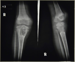
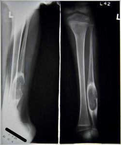
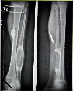
Further skeletal changes are observed in the x-ray report notes from September 1939, Figures 6 and 7:

11/9/39 –             Leg – large cavity in fibula L and in head of R. tibia

15/9/39 –             Left fibula large focus

     Right tibia large focus passing through into epiphysis.

Combined with this the medical notes indicate that a sinus developed on the left ankle and another on the right tibia during the same period with a further sinus developing in November 1939.


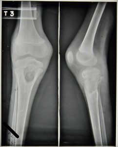
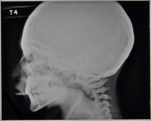
This patient was transferred from Stannington in February 1940 to a local hospital in West Hartlepool, his home town, as showing No Medical Improvement and a final diagnosis of TB Bones and Joints and old lung lesion.  His final x-ray report, see Figures 8-12, dated 27th February 1940, reads:

Large cavity head of R.tibia & sequestrum seem smaller than 11/9/39

Elbow –Improved

Fibula – large cavity little change.

Skull – little seen

Chest – L.apex clearer much mottling’

HOSP-STAN-07-01-02-91_18
FIGURE 8: HOSP-STAN-07-01-02-91_18
HOSP-STAN-07-01-02-91_17
FIGURE 9: HOSP-STAN-07-01-02-91_17


HOSP-STAN-07-01-02-91_03
FIGURE 10: HOSP-STAN-07-01-02-91_03
HOSP-STAN-07-01-02-91_04
FIGURE 11: HOSP-STAN-07-01-02-91_04
HOSP-STAN-07-01-02-91_05
FIGURE 12: HOSP-STAN-07-01-02-91_05


The multifocal nature of this patient coupled with comments throughout the notes on possible non-TB origin is suggestive of a potential differential diagnosis. Any further comments based upon the information provided and radiographic images would be welcomed.