🎄Christmas Opening Hours for 2024🎄
CLOSED between 4pm Friday 20th December 2024 and 10am Thusday January 2nd 2025
Ordinary opening hours apply before and after this period.

Surgical Procedures – Osteotomy

Patient 91/10 was a 15 year old boy admitted to Stannington on 31st October 1941, a letter attached to his discharge report alludes to him having been transferred to Stannington from Newcastle. His initial examination notes reveal he had some tightness over the abdominal area and the right hip was in a state of 45° flexion with some adduction, slight exterior rotation and wasted muscle. Signs of two previous sinuses were also present on the right thigh. The patient had previously been admitted to Stannington from August 1936 until February 1939 when he was discharged in a splint which he wore until the age of 13 prior to his second admission and his hip became flexed; unfortunately we do not hold records for patients this early on and as such have no further details about his previous stay. Figures 1 and 2 are two of the radiographs of his first stay in Stannington, the first showing some tuberculous activity in the lungs and the second showing tuberculous involvement in the right hip joint.
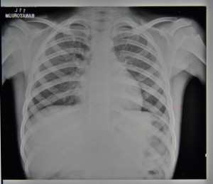
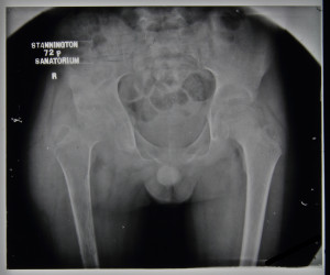
FIGURE 1: HOSP-STAN-07-01-02-686_12
FIGURE 2: HOSP-STAN-07-01-02-686_10


This boy was diagnosed with 3rd stage TB of the right hip and was put forward for a surgical procedure known as an osteotomy. An osteotomy, known as ‘bone cutting’, is a procedure whereby the bone near a damaged joint is cut to realign load bearing surfaces. In the case of patient 91/10 a right femoral osteotomy was performed on the proximal femur to better align it with the acetabulum (socket) of the pelvis, most likely to improve functionality of the limb.

Initial notes on this patient from early 1942 are vague referring to the levels of flexion and adduction in the hip joint but little else. The x-ray report prior to the osteotomy note that no infection is active in the lungs and that there was firm ankyloses in the hip. The osteotomy took place on 13th February 1942.
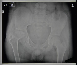
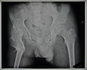
 

FIGURE 3: HOSP-STAN-07-01-02-686_05
FIGURE 4: HOSP-STAN-07-01-02-686_04


Figures 3 and 4, respectively, show the patient’s condition before and after the osteotomy was carried out. Figure 3 shows tuberculous infection in the hip joint, affecting the femoral head with some acetabulum involvement. The right hip appears on a slant in comparison to the left sitting higher up, this could potentially have caused the patient to have limp. Figure 4, taken on 14th April 1942, shows the results of the osteotomy, where the femoral head has been repositioned with the aim of realigning the hip joint.

Following the procedure, recovery appears to have gone relatively smoothly. A plaster splint was applied to the patient on 3rd March 1942 where it was noted that the femur had ‘40° abduction – no change’.

No complications are outlined within the notes and the x-ray reports state that there was:

                        ‘14/4/42 –            Good bony union after osteotomy. Position satisfactory.

                        17/7/42 –            Chest no change.

                                                   Hip – osteotomy satisfactory good ankylosis.’


By June 1942 the patient was capable of standing and putting weight on the right leg and is noted to be walking well by mid-July, meriting the osteotomy a great success. The patient was discharged on the 24th July 1942 as quiescent after a 38 week stay at Stannington. The final surgical outcome can be seen in Figure 5.
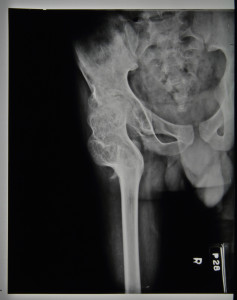
FIGURE 5: HOSP-STAN-07-01-02-686_14


To see more radiographic images from Stannington, have a look at our Flickr Stream https://www.flickr.com/photos/99322319@N07/sets/72157648833066476

Sources:

Tuli, S.M (2004) Tuberculosis of the Skeletal System: Bones, Joints, Spine and Bursal Sheaths. Jaypee Brothers Publishers.

Memories of Stannington

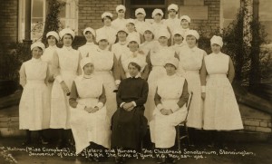
The records of Stannington Sanatorium came to Northumberland Archives in the 1980s and at this point a former patient who was in the sanatorium from 1930-1936 wrote to the then Deputy County Archivist sharing his memories of his time in Stannington:


Dear Sir

What a joy and lovely surprise to receive your letter & brochure about Stannington Sanatorium!  I was overcome and close to tears, as having spent nearly 5 ½ years there as a child, & in my formative years Stannington has meant so much to me.  I had a TB knee my parents took me to my Dr. who confirmed it & sent me to Newcastle Infirmary for treatment.  Leg put in Paris of Plaster, did no good to my leg, so I was sent to Stannington feeling terribly homesick & cried every night to go home, however I settled down to a long sojourn & treatment.

Visiting day was once every two months!  My mother, a lovely soul, used to send me books & comics every week.  The Magnet, The Gem, Adventure & Hotspur etc.  How I looked forward to receiving them and a five shilling postal order to buy things at the tuck shop.  Those were the days of hospitals with matrons, sisters, ward sisters, a lady almoner, no unions, but very dedicated nurses and staff, people who loved what they were doing.  Oh yes ward maids who loved us children, the matron was strict but understanding & a comfort too, the nurses used to cuddle us and show us a lot of affection.  I loved it all!

HOSP/STAN/11/1/65
HOSP/STAN/11/1/65

I received good food, had a spoonful of Virol and Numol twice a day, Christmas was special, artists from the Newcastle Empire & Palace came to entertain us, nurses sang carols.  I feel sure we became their children to love and care for I received many kisses from them, the nearness of them as they carried us off to the bathroom was sweet & to a child they became our mothers bless them.  Yes I owe Stannington a lot for my education & way of life because remember it was the thirties & people were hungry and home, no work for men and lots of pawn shops open.  But there was no violence, or muggings as they call it and we could walk the streets at without fear & folk were caring and kind, what a sad state of affairs today Sir.

I was born and bred in Wallsend served my apprenticeship as a joiner at Swan Hunters very strict too.  I had a happy time as a youth, used to dance at the Oxford Galleries, drink at the Pineapple Grill & go every Saturday to the Empire to see visiting bands such as Roy Fox, Harry Roy, Lew Stone etc. and also to a 4 hour show at the Paramount Cinema complete with organ and stage show all for half a crown.  Oh happy days!

I am returning in three weeks’ time I remember a lot of my little friends who died in Stannington while I have been blessed with a good working life, a family & good health.  I am a true Geordie in my nature and spirit I am full of nostalgia for Stannington and Newcastle and those people who were true and dedicated to their work who gave a little boy from Wallsend on Tyne lots of love and care and put me on my feet again.  I have worked out since, in my memory summers were long, people were nice and one never grew old.

Thank you

[HOSP/STAN/13/5]

Miliary TB

Miliary spread is a pathological process involving the widespread dissemination of the tubercle bacilli but the manifestations of this can vary widely depending on factors such as the speed of the spread and the individual’s ability to inhibit further multiplication of the organisms in other areas of the body.  Miliary tuberculosis is recognised clinically in patients where there is radiographic evidence of tuberculous lesions of the same age distributed evenly throughout all areas of the lung.  This process is most likely to occur soon after the initial infection and is also more common in children under 5 than it is in older children.


It is not uncommon to see miliary tuberculosis develop further into tuberculous meningitis.  Whilst on the whole the death rate in Stannington was relatively low, in the pre-antibiotic era (pre-1947) it is noticeable that a significant number of fatalities are as a result of either miliary TB or TB meningitis.  The introduction of effective drug therapies altered this situation greatly and the prospects for these patients after this point improved significantly.


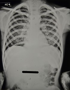
Patient 3/1947 was a 12 year old boy from Lemington-on-Tyne who was admitted to Stannington in January 1947 diagnosed with miliary tuberculosis.  A report on x-ray films taken in December 1946 prior to his admission describes extensive mottled shadowing across both lungs with hilar shadows much enlarged.  The Northumberland County medical officer of health that refers the boy to Stannington gives the following report:

‘States no cough.  Mother says he thinks he gets a bit short of breath at times, and that he has definitely lost weight.  On examination, slight cyanotic tinge; afebrile, pulse 108.  General condition satisfactory (amazing in view of films).  Little made out in chest apart from slight impairment of the air entry at both bases.  Mass of glands at right side of neck.’


Figure 1 is a chest x-ray taken the day after his admission and the report on it simply reads, ‘extensive bilateral miliary spread’.  The extensive mottled ‘snowstorm’ effect is indicative of miliary TB.  Strict bed rest is ordered and at this point he also has an enlarged gland at the angle of the jaw on the right side for which UV light treatment is prescribed.  Over the coming months the abscess on the jaw is described as discharging freely with brownish pus aspirated from it in June 1947.

Figure 1 - HOSP/STAN/7/1/2/1371_84
Figure 1 – HOSP/STAN/7/1/2/1371_84
09 Jan 1947
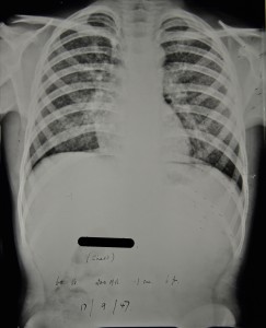
Figure 2 - HOSP/STAN/7/1/2/1371_05
Figure 2 – HOSP/STAN/7/1/2/1371_05
17 Sept 1947


In September 1947 the patient’s doctor suggests that he would be a suitable candidate for streptomycin treatment, which had only recently been introduced at this point.  However, having discussed the case further it was decided that he was not suitable as at this point in time streptomycin was being used for very early cases only and patient 3/1947 by now had a long history of TB and was doing very well without it.  Figure 2, is an x-ray taken around the time streptomycin treatment was being discussed and the report reads, ‘X-ray shows a little improvement.  Each individual lesion is smaller.’


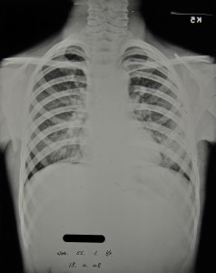
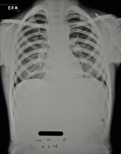
Two months later in November 1947 his condition deteriorates a little and he begins to lose weight and so is again put forward for streptomycin ‘if any available.’  Whilst the attending doctor continues to push for streptomycin over the coming months it is not until November 1948 that the patient receives any.  There are continuing disputes as to whether he is a suitable candidate.  During this time his general condition fluctuates with periods of weight gain and weight loss and x-rays from April and June 1948 show some improvements, figures 3 and 4 respectively.

Figure 3 - HOSP/STAN/7/1/2/1371_09
Figure 3 – HOSP/STAN/7/1/2/1371_09
13 April 1948
‘Amazing improvement since last x-ray 3 months ago. The military lesions now appear to be resolving: the apices are almost clear. There is now a more homogenous opacity in the left lower lobe.’
Figure 4 - HOSP/STAN/7/1/2/1371_10
Figure 4 – HOSP/STAN/7/1/2/1371_10
04 June 1948
‘still some mottling at the bases, the apices are clear. The more homogenous density at the L base is getting smaller. Azygos lobe on right side.’


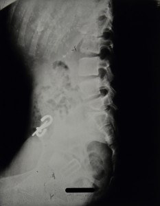
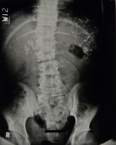
Ultimately the catalyst leading to the decision being made for streptomycin treatment to be commenced appears to be the fact that in June 1948 a swelling over the lower dorsal spine is identified and it is apparent that the tuberculous infection has spread further.  It is clear from radiographic evidence in 1948 that there are three spinal lesions: one in the 5th and 6th dorsal vertebrae causing some kyphosis; another affecting the 1st and 3rd lumbar vertebrae causing some deformity; and a final one in the inferior and anterior part of the body of the 5th lumbar vertebrae with some destruction.


All streptomycin treatment is discontinued by May 1949 when significant improvements in his chest are seen and treatment of the spinal lesions is continued with braces and plaster casts.  He is eventually discharged in July 1950 wearing a spinal brace and continues to be seen by the out-patients’ service until April 1954 when an abscess in the left iliac fossa leads to him being considered for admission to the Queen Elizabeth Hospital.

Figure 5 - HOSP/STAN/7/1/2/1371_18
Figure 5 – HOSP/STAN/7/1/2/1371_18
Spinal X-ray, 17 Jan 1950, calcifications in the lungs also evident.
Figure 6 - HOSP/STAN/7/1/2/1371_85
Figure 6 – HOSP/STAN/7/1/2/1371_85
Spinal X-ray, 10 July 1950, 4 days before discharge
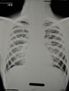
Figure 7 - HOSP/STAN/7/1/2/1371_14 Chest X-ray, 17 Feb 1950, also showing calcifications in neck glands.
Figure 7 – HOSP/STAN/7/1/2/1371_14
Chest X-ray, 17 Feb 1950, also showing calcifications in neck glands.


Sources:

MILLER, F. J. W, SEAL, R. M. E, and TAYLOR, M. D. (1963) Tuberculosis in Children, J & A Churchill Ltd.