|
X-ray Plant - History
X-rays and their potential for medical imaging were discovered in 1895 by the German physicist Wilhelm Conrad Röntgen. They serve as a great diagnostic tool in the field of medicine and as far as TB is concerned allow for much greater accuracy in its diagnosis and the identification of the stage the disease is at. The routine screening of individuals who had been in contact with someone known to be infectious was carried out by tuberculosis dispensaries and became quite commonplace by the 1940s. By catching and identifying the disease early the prospect of a successful outcome from treatment was much greater.
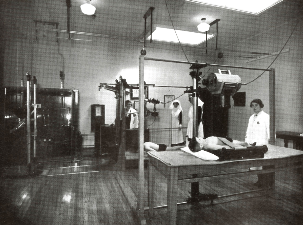
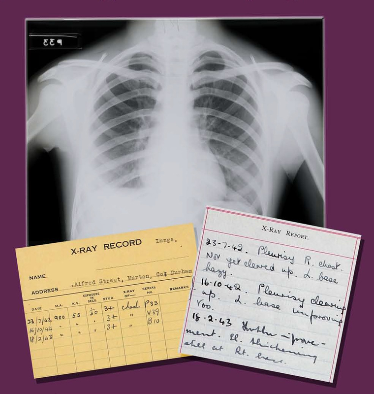
An x-ray plant was first installed in Stannington in 1920 and all patients would have been routinely radiographed upon admission and throughout their treatment. The 14,660 radiographs within the collection are grouped by patient into 2,243 sets. Whilst some patients might only have had one x-ray taken others have as many as 80. They can all be matched up to the relevant patient case file in which it is common to find x-ray reports written by the child's physician explaining what can be seen in the image.
The regular x-raying of each child meant that the medical staff could closely monitor how well they were responding to the treatment and if indeed the treatment was working at all. This was particularly useful in lung collapse therapies, such as artificial pneumothorax, that were used to treat pulmonary TB as the x-rays would illustrate exactly how well the lung was collapsing. The possibility of the disease having spread to other parts of the body could also be monitored and the stage the disease was at more easily ascertained.
|