🎄Christmas Opening Hours for 2024🎄
CLOSED between 4pm Friday 20th December 2024 and 10am Thusday January 2nd 2025
Ordinary opening hours apply before and after this period.

Surgical Procedures – Artificial Pneumothorax

Pulmonary tuberculosis is by far the most common manifestation of TB witnessed throughout the Stannington records.  Prior to the development and use of any effective antibiotic treatments the most common form of intervention was the induction of an artificial pneumothorax.  Many of the different treatments employed to treat TB of all types at this time were based on the principles of resting and isolating the affected area, and the thinking behind artificial pneumothorax treatment was no different.


A needle would be inserted through the chest wall to allow for the insertion of air into the pleural cavity.  The amount of air inserted would depend on the size of the patient as well as how much the physician in charge though the patient could realistically manage in one go and how quickly they wished the lung to collapse.  Once inserted the pressure from the air would force the lung to collapse in on itself and to cease functioning properly.  The entire lung would not necessarily be collapsed at once, either because it wasn’t necessary for treatment or because fibrotic adhesions between the lung and the chest wall as a result of the disease prevented it from doing so.  Where only part of the lung was affected it would not be desirable to collapse the whole lung and in such instances just one lobe might be collapse.  Bilateral artificial pneumothorax was also a possibility, whereby part of both lungs would be collapsed at the same time.  A state of collapse could be maintained for a period of months or even years and required the patient to undergo regular refills of air in order to do so.


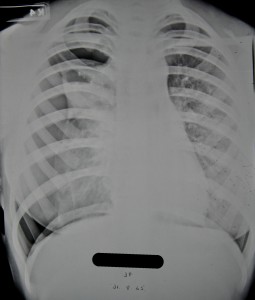
A great number of radiographic illustrations of the progression of a collapse are available in the Stannington collection.  One patient, 2/1946, has a large amount of radiographs taken over a period of two years which demonstrate the change in the lung from admission and through the progressive stages of lung collapse.


Patient 2/1946 was female an age15 when she was admitted to Stannington on 21 June 1945 with pulmonary TB stage 3, at which point her sputum tested positive for TB also.  A report on an x-ray taken pre-admission reads:

‘Right lung shows several active foci beginning to coalesce.  There is extensive infiltration in the upper zone & suspicious blotchy areas in the middle zone.  A small calcified opacity in the right lower zone.  The left lung shows infiltration in the middle zone.  The upper zone and apex are clear.  Early active foci are noticeable in both lungs in the affected areas.’

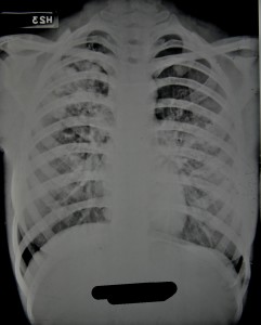
Figure 1 was the first x-ray taken after admission on 25 June 1945 being three weeks later than the one reported above.  Observations on this x-ray note:

‘Scattered foci in right upper zone.  One definite cavity.  Increased bronchial marking at both bases.’
HOSP/STAN/7/1/2/1057_22 25 June 1945
Figure 1 – HOSP/STAN/7/1/2/1057_22
25 June 1945
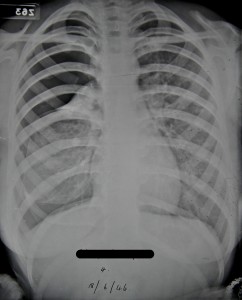
HOSP/STAN/7/1/2/1057_18 31 Aug 1945
Figure 2 – HOSP/STAN/7/1/2/1057_18
31 Aug 1945


It was quickly decided that and artificial pneumothorax should be induced on the right side and this took place on 16 Aug 1945. Figure 2 taken later on that month shows the initial results of the artificial pneumothorax.  The black area along the lateral side of the right lung is evidence of the air that has been inserted and the lung has begun to compress.


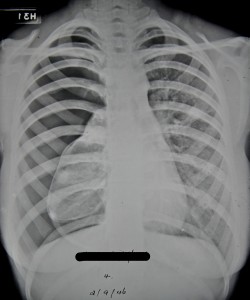
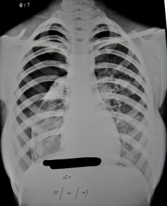
The collapse was maintained well into 1947 which involved her having refills of air every two weeks throughout this period.  For the first three months she received refills of 200-300ccs of air at a time, progressing to 400ccs the month after, and then eventually 500-600ccs at a time.  Figures 3-6 show the progression of the artificial pneumothorax as more air is inserted and the lung collapses further.  Over time we can see that the cavity in the right mid zone collapses and closes, one of the main aims of the treatment.  In early June 1946 a procedure was performed to divide adhesions between the lung and the chest wall which allowed the collapse to progress further.  She was discharged in June 1947 with her condition described as improved.
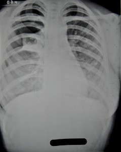
 

Figure 3 - HOSP/STAN/7/1/2/1057_23 17 Jan 1946
Figure 3 – HOSP/STAN/7/1/2/1057_23
17 Jan 1946
Figure 4 - HOSP/STAN/7/1/2/1057_09 18 June 1946
Figure 4 – HOSP/STAN/7/1/2/1057_09
18 June 1946


Figure 5 - HOSP/STAN/7/1/2/1057_10 2 Sept 1946
Figure 5 – HOSP/STAN/7/1/2/1057_10
2 Sept 1946
Figure 6 - HOSP/STAN/7/1/2/1057_27 15 April 1947
Figure 6 – HOSP/STAN/7/1/2/1057_27
15 April 1947


Patient 90/38, An Unconfirmed Diagnosis

Amongst the patients admitted to Stannington Sanatorium there are a number for which following admission doctors decide that their condition for whatever reason is non-tuberculous.  Differential diagnoses can vary from bronchiectasis and asthma in those suspected of having pulmonary TB to Perthes’ Disease in those suspected of having TB of the hip.  One patient who ultimately appears not to have TB is patient 90/38, a 17 ½ year old girl presenting with strong neurological symptoms, although no definite conclusions seem to be drawn on what the cause might be.


Admitted on 12 Sept 1941, she is one of the very few private patients and also one of the oldest.  The diagnosis given at the top of her file is ‘Non-TB, query bone tumour spine and skull’.  She had been suffering from symptoms for a year prior to admission and reports immediately following admission state ‘Lower thoracic curvature, no active angular deformity.  Not tuberculous’.


The first x-rays of her spine are taken the day after admission and here the report reads:

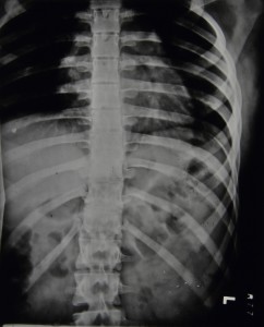
Marked irregularity of epiphyses in lower thoracic region.  Some wedging of bodies of 9th and 10th dorsal vertebrae.  Edges of bone are irregular & ossification is either incomplete or of poor quality.

                Diagnosis: Epiphysitis of thoracic region, probably not tubercular

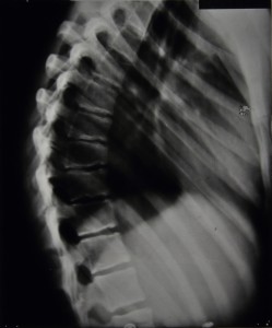
Over the coming months further spinal x-rays and their corresponding reports do not suggest any significant worsening of the spinal wedging nor any great improvements.  The final report indicates that 5 vertebrae are affected with the 9th and 10th being the worst.  Figures 1 and 2 are examples of some of the spinal x-rays that were taken.


HOSP/STAN/7/1/2/651_25
Figure 1 – HOSP/STAN/7/1/2/651_25
HOSP/STAN/7/1/2/651_03
Figure 2 – HOSP/STAN/7/1/2/651_03


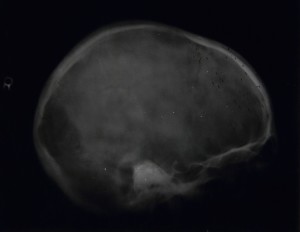
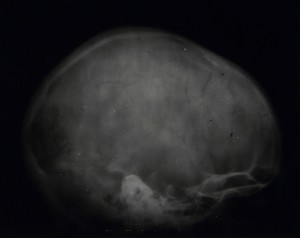
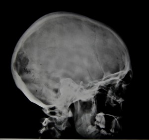
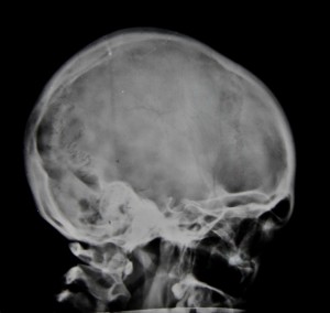
In addition, x-rays were taken of her arms, forearms, pelvis, femora, and legs, all of which were clear.  There are also 7 x-rays taken of the skull, 4 of which can be seen in figures 3-6. Reports on the skull x-rays read as follows:

9/12/1941: Skull, localised deficiency of internal table to left of midline – lying over leg area.

19/3/1942: Outline of internal table broken for about 1” in anterior-parietal region. 

14/5/1942:  Rarefaction appears to be falling in.  Outline more normal.  Break still about 1”. 

Figure 3 - HOSP/STAN/7/1/2/651_23
Figure 3 – HOSP/STAN/7/1/2/651_23
Figure 4 - HOSP/STAN/7/1/2/651_14
Figure 4 – HOSP/STAN/7/1/2/651_14


Figure 5 - HOSP/STAN/7/1/2/651_21
Figure 5 – HOSP/STAN/7/1/2/651_21


Figure 6 - HOSP/STAN/7/1/2/651_04
Figure 6 – HOSP/STAN/7/1/2/651_04


Her file also contains quite detailed reports on other tests carried out and her general condition during her stay.  In November 1941 reports are made of signs of mental disturbance and that she ‘will not speak to anyone and only laughs or cries when spoken to’.  She is also experiencing some incontinence and has a history of incontinence between the ages of 8 and 14.  She has bilateral ankle clonus and a positive Babinski test, more marked on the right.  Two days later the report reads as follows:

Spasticity lower limbs.  KJs +.  Bilateral ankle clonus.  Plantar Reflex? – probably flexor.  Sensation apparently normal.  Pupils reacting normally.  Eye movements, other cranial nerves & field of vision-apparently normal but patient unresponsive & difficult to examine.

She says she feels miserable & that everyone thinks she is silly, & that she has been like this before.

Still some incontinence.


At the end of November 1941 it is noted that there is a white patch in the centre of the optic discs and that the disc edges are blurred, still some spasticity, slight clonus, sluggish Babinski, normal co-ordination, normal mental condition, and occasionally experiences some frontal headaches.  In March of 1942 a Wassermann Test comes back negative and she is eventually discharged on 16th May 1942.

If anyone can offer any further opinions on the possible causes of her condition please feel free to add your comments below.

Tuberculous-Arthritis of the Knee

Tuberculosis of the bones and joints affected several key areas of the body, and is well documented amongst the Stannington records. Of these the knee is one of the more frequently noted areas of infection. Immobilisation by plaster cast was the most common form of treatment for this type of tuberculosis, although some more severe cases were put forward for surgical intervention.

Tuberculous arthritis characteristically affects only one joint, predominantly a weight-bearing joint such as the spine, hip or knee. It is transferred by haematogenous spread from a location of primary infection, most commonly the lungs. Initial symptoms often include synovitis or inflammation of the soft tissue in addition to joint effusion, where there is an increase in the fluid within the joint. These preliminary symptoms progress into arthritis over a period of time, although radiographic findings only begin to occur after three or four weeks. Ultimately, untreated tuberculous arthritis will lead to demineralisation, erosion and joint destruction.

Case Study

HOSP-STAN-07-01-02-1344_02
HOSP-STAN-07-01-02-1344_04


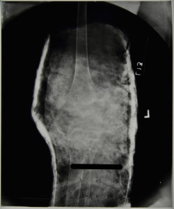
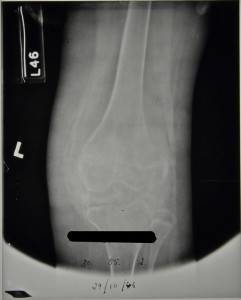
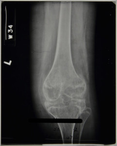
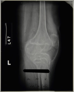
Patient 358/1946 was admitted to Stannington Sanatorium in October 1946 with tuberculous arthritis of the left knee. The patient notes detail that on admittance there was radiographic evidence of destructive lesions already identifiable, however, the first radiographs taken of the individual are of poor exposure or whilst the individual was in plaster cast, so identification is challenging.

The radiographs from February 1947 show the bony anomalies to the knee joint clearly. There is a significant reduction in joint space between the femur and the tibia. The distal epiphysis of the femur shows severe displacement, having moved towards the posterior. Similar displacement can be seen on the proximal tibia to a slightly lesser degree. The patient notes at this stage indicate no change from time of admittance that two sinuses were present above the patella and that immobilisation of the knee was to continue.


HOSP-STAN-07-01-02-1344_05
HOSP-STAN-07-01-02-1344_06


In August 1947, an examination by the visiting physician describes: 

‘Complete disorganisation of the joint. Less decalcification and bony trabeculae are beginning to show.

Fusion of the joint is not complete and there is still some heat.

To be put in plaster for three months’

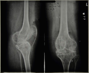
Changes in the radiographic images between February 1947 and June 1948, when the patient is discharged, are minimal. In December 1947 the physician stated in the patient’s notes:

‘No change in appearance.

There is not complete bony ankyloses of the knee but movement is negligible.

A sinus on the front of the knee which is covered by a scab, is not at present discharging’

There is little or no heat in the knee.

For Thomas’ walking knee splint, patton and crutches.’

No further changes were noted at this stage with the radiographic image below, dated to December 1947, revealing gross anatomical destruction of the knee joint to have taken place and there is no remaining joint space. The striation pattern across the epiphysis and metaphysis of both the femur and tibia is likely to be the result of cartilage destruction and bone degeneration causing porosity in the bones.


HOSP-STAN-07-01-02-1344_13

Patient 358/1946 was discharged in June 1948 but according to their patient notes returned twice as an out-patient and was seen a further two times at the Sanderson Orthopaedic Hospital, Gosforth.


For a case study on the surgical interventions used in tuberculosis of the hip, see earlier post of 08/12/2014

Further radiographic images can be seen on Flickr at https://www.flickr.com/photos/99322319@N07/sets/72157648833066476/


Sources

Albuquerque-Jonathan, G (2006). Atypical tuberculosis of the knee joint. South African Journal of Radiology p.28.

Arthanari, S; Yusuf, S and Nisar, M (2008). Tuberculosis of the Knee Complicating Seronegative Arthritis. Journal of Rheumatology: http://www.jrheum.com/subscribers/08/06/1227.html