Patient 81/39 – A Questionable Diagnosis?

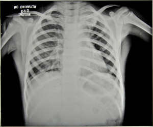
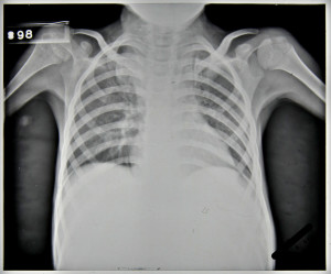
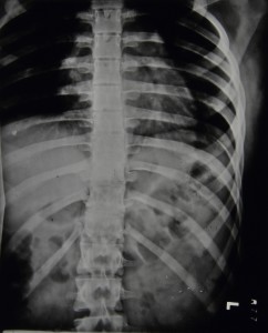
Patient 81/39, a five year old boy, was admitted to Stannington in December 1937 due to ailing health following a two month period in bed suffering from mumps. He had developed a cough, was easily tired and was losing weight. The initial x-ray reports detail a blocked apex in the left lung and marked mottling in the right lung leading to an initial diagnosis of Pulmonary TB, Figure 1. However, following his admission further symptoms started to manifest themselves which indicated that the diagnosis of this patient was more complex than it was initially considered to be.
HOSP-STAN-07-01-02-91_01
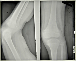
FIGURE 2: HOSP-STAN-07-01-02-91_01
HOSP-STAN-07-01-02-91_15
FIGURE 1: HOSP-STAN-07-01-02-91_15


HOSP-STAN-07-01-02-91_14
FIGURE 3: HOSP-STAN-07-01-02-91_14


In April 1938, it was noted the patient had two subcutaneous abscesses on the iliac crest and the knee. A sample of the mucus taken from the abscess on the hip was sent for bacteriological examination. Results of this testing were as follows:

scanty pus cells and much granular debris. No definite organisms seen and tubercle bacilli not found.’

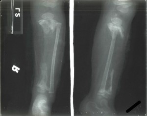
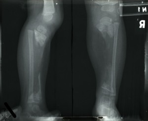
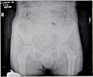
Furthermore, periostitis was noted in the upper end of the ulna which ‘appears septic’ but was regarded as being non-tuberculous. The patient still suffered with a cough but sputum tests were negative and notes state that no tuberculosis was seen. At this stage the x-ray report indicates that no bone lesions are seen in either the leg or the iliac crest, Figures 2 and 3.

Throughout the rest of 1938, the patient’s condition is very variable. An additional abscess is noted in the lumbar region with slight discharge and the apex of the left lung becomes more blocked with the lower lobe of the right lung being described as having been ‘studded with deposits’, however, the sinuses in the thigh and gluteus region are healed.
HOSP-STAN-07-01-02-91_19
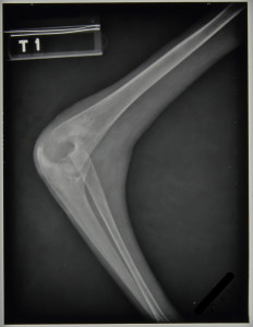
FIGURE 4: HOSP-STAN-07-01-02-91_19
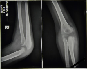
FIGURE 5: HOSP-STAN-07-01-02-91_13


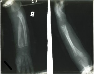
The main focus of the notes centre upon the right elbow which, in September 1938, was described as being very active with discharging abscesses; periostitis was greatly increased in the ulna and also present in the humerus with the joint being ‘badly involved’, see Figure 4. In November 1938 large sequestrum was removed from the elbow, at this time all lesions were considered very active. The elbow continued to be active with an increasing number of ulcers noted to have appeared; a maximum of four seen in February 1939 including one in the right cubital fossa which is incised to produce ‘copious…pus’, Figure 5.

X-ray reports from September 1939 read as follows:

11/9/39 –              Ulna hollowed out to cavity

                            Radius dislocated upward & forward

                            Lower end humerus eroded & partly destroyed.

15/9/39 –            Ulna – upper end partially destroyed, disorganisation of elbow joint’

No further comment is made regarding a diagnosis of tuberculosis in the elbow.


In addition to ongoing changes in the elbow an abscess appeared on the right mastoid, which was opened and drained in October 1938 and is noted to have become less active by November 1938. However, this abscess continued to open throughout the patient’s stay at Stannington and is often referred to as ‘discharging freely,’ with a diminishment in its activeness finally being noted in October 1939.
HOSP-STAN-07-01-02-91_09
FIGURE 6: HOSP-STAN-07-01-02-91_09
HOSP-STAN-07-01-02-91_02
FIGURE 7: HOSP-STAN-07-01-02-91_02


Further skeletal changes are observed in the x-ray report notes from September 1939, Figures 6 and 7:

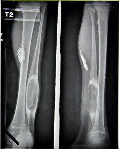
11/9/39 –             Leg – large cavity in fibula L and in head of R. tibia

15/9/39 –             Left fibula large focus

     Right tibia large focus passing through into epiphysis.

Combined with this the medical notes indicate that a sinus developed on the left ankle and another on the right tibia during the same period with a further sinus developing in November 1939.


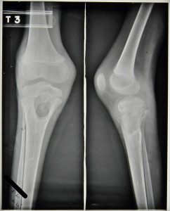
This patient was transferred from Stannington in February 1940 to a local hospital in West Hartlepool, his home town, as showing No Medical Improvement and a final diagnosis of TB Bones and Joints and old lung lesion.  His final x-ray report, see Figures 8-12, dated 27th February 1940, reads:

Large cavity head of R.tibia & sequestrum seem smaller than 11/9/39

Elbow –Improved

Fibula – large cavity little change.

Skull – little seen

Chest – L.apex clearer much mottling’
HOSP-STAN-07-01-02-91_18
FIGURE 8: HOSP-STAN-07-01-02-91_18
HOSP-STAN-07-01-02-91_17
FIGURE 9: HOSP-STAN-07-01-02-91_17


HOSP-STAN-07-01-02-91_03
FIGURE 10: HOSP-STAN-07-01-02-91_03
HOSP-STAN-07-01-02-91_04
FIGURE 11: HOSP-STAN-07-01-02-91_04
HOSP-STAN-07-01-02-91_05
FIGURE 12: HOSP-STAN-07-01-02-91_05


The multifocal nature of this patient coupled with comments throughout the notes on possible non-TB origin is suggestive of a potential differential diagnosis. Any further comments based upon the information provided and radiographic images would be welcomed.

Patient 90/38, An Unconfirmed Diagnosis

Amongst the patients admitted to Stannington Sanatorium there are a number for which following admission doctors decide that their condition for whatever reason is non-tuberculous.  Differential diagnoses can vary from bronchiectasis and asthma in those suspected of having pulmonary TB to Perthes’ Disease in those suspected of having TB of the hip.  One patient who ultimately appears not to have TB is patient 90/38, a 17 ½ year old girl presenting with strong neurological symptoms, although no definite conclusions seem to be drawn on what the cause might be.


Admitted on 12 Sept 1941, she is one of the very few private patients and also one of the oldest.  The diagnosis given at the top of her file is ‘Non-TB, query bone tumour spine and skull’.  She had been suffering from symptoms for a year prior to admission and reports immediately following admission state ‘Lower thoracic curvature, no active angular deformity.  Not tuberculous’.


The first x-rays of her spine are taken the day after admission and here the report reads:

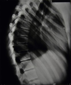
Marked irregularity of epiphyses in lower thoracic region.  Some wedging of bodies of 9th and 10th dorsal vertebrae.  Edges of bone are irregular & ossification is either incomplete or of poor quality.

                Diagnosis: Epiphysitis of thoracic region, probably not tubercular

Over the coming months further spinal x-rays and their corresponding reports do not suggest any significant worsening of the spinal wedging nor any great improvements.  The final report indicates that 5 vertebrae are affected with the 9th and 10th being the worst.  Figures 1 and 2 are examples of some of the spinal x-rays that were taken.


HOSP/STAN/7/1/2/651_25
Figure 1 – HOSP/STAN/7/1/2/651_25
HOSP/STAN/7/1/2/651_03
Figure 2 – HOSP/STAN/7/1/2/651_03


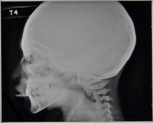
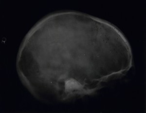
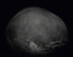
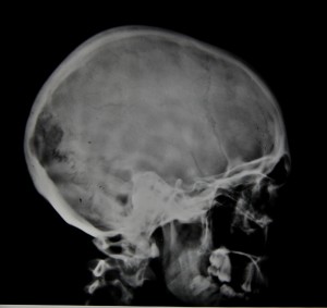
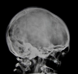
In addition, x-rays were taken of her arms, forearms, pelvis, femora, and legs, all of which were clear.  There are also 7 x-rays taken of the skull, 4 of which can be seen in figures 3-6. Reports on the skull x-rays read as follows:

9/12/1941: Skull, localised deficiency of internal table to left of midline – lying over leg area.

19/3/1942: Outline of internal table broken for about 1” in anterior-parietal region. 

14/5/1942:  Rarefaction appears to be falling in.  Outline more normal.  Break still about 1”. 

Figure 3 - HOSP/STAN/7/1/2/651_23
Figure 3 – HOSP/STAN/7/1/2/651_23
Figure 4 - HOSP/STAN/7/1/2/651_14
Figure 4 – HOSP/STAN/7/1/2/651_14


Figure 5 - HOSP/STAN/7/1/2/651_21
Figure 5 – HOSP/STAN/7/1/2/651_21


Figure 6 - HOSP/STAN/7/1/2/651_04
Figure 6 – HOSP/STAN/7/1/2/651_04


Her file also contains quite detailed reports on other tests carried out and her general condition during her stay.  In November 1941 reports are made of signs of mental disturbance and that she ‘will not speak to anyone and only laughs or cries when spoken to’.  She is also experiencing some incontinence and has a history of incontinence between the ages of 8 and 14.  She has bilateral ankle clonus and a positive Babinski test, more marked on the right.  Two days later the report reads as follows:

Spasticity lower limbs.  KJs +.  Bilateral ankle clonus.  Plantar Reflex? – probably flexor.  Sensation apparently normal.  Pupils reacting normally.  Eye movements, other cranial nerves & field of vision-apparently normal but patient unresponsive & difficult to examine.

She says she feels miserable & that everyone thinks she is silly, & that she has been like this before.

Still some incontinence.


At the end of November 1941 it is noted that there is a white patch in the centre of the optic discs and that the disc edges are blurred, still some spasticity, slight clonus, sluggish Babinski, normal co-ordination, normal mental condition, and occasionally experiences some frontal headaches.  In March of 1942 a Wassermann Test comes back negative and she is eventually discharged on 16th May 1942.

If anyone can offer any further opinions on the possible causes of her condition please feel free to add your comments below.

Unreported X-Rays

Amongst the 2242 sets of patient radiographs we hold there are 55 for which we do not have any corresponding patient case notes.  Within this group of 55 there is one patient in particular that stands out as the radiographs that survive for him are the earliest original radiographs we hold with the others all being on microfiche.  The other 54 also have their patient numbers clearly marked on the radiographs and references to them can be found amongst the other records in the collection, for example admission registers and treatment registers.


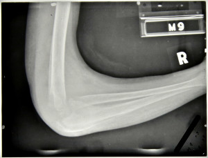
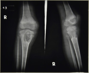
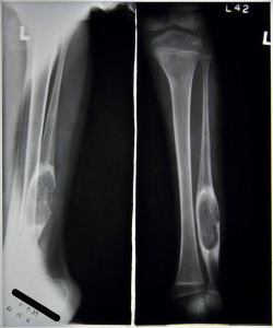
All we know about this patient is his name and the date on which the x-rays were taken.  The three x-rays were taken in September and October of 1939 and show the boy’s right arm and right leg.  Judging from his size we are assuming he was relatively young and was evidently suffering from disease of the right tibia and right forearm.  There are no later re-admissions for this patient and so we are assuming that he either recovered and was discharged with no later relapse or that he died.  We have the sanatorium school records for the period as well and there is no mention of him there, so again we can assume he was either below school age or too ill to attend.  The only other contemporary records we have where we might expect to find him are the register of operations and the register of splints and appliances.  Again there is no reference in either of these confirming he did not receive any surgical treatment during his stay and was not issued with a splint.


HOSP-STAN-7-1-2-2-1 HOSP-STAN-7-1-2-2-2

HOSP-STAN-7-1-2-2-3Click on the images to enlarge


If you can offer any additional details about this patient from his radiographs please add them to the comments below.