🎄Christmas Opening Hours for 2024🎄
CLOSED between 4pm Friday 20th December 2024 and 10am Thusday January 2nd 2025
Ordinary opening hours apply before and after this period.

Tuberculous-Arthritis of the Knee

Tuberculosis of the bones and joints affected several key areas of the body, and is well documented amongst the Stannington records. Of these the knee is one of the more frequently noted areas of infection. Immobilisation by plaster cast was the most common form of treatment for this type of tuberculosis, although some more severe cases were put forward for surgical intervention.

Tuberculous arthritis characteristically affects only one joint, predominantly a weight-bearing joint such as the spine, hip or knee. It is transferred by haematogenous spread from a location of primary infection, most commonly the lungs. Initial symptoms often include synovitis or inflammation of the soft tissue in addition to joint effusion, where there is an increase in the fluid within the joint. These preliminary symptoms progress into arthritis over a period of time, although radiographic findings only begin to occur after three or four weeks. Ultimately, untreated tuberculous arthritis will lead to demineralisation, erosion and joint destruction.

Case Study
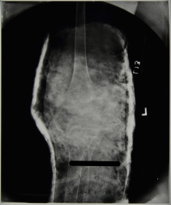
HOSP-STAN-07-01-02-1344_02
HOSP-STAN-07-01-02-1344_04


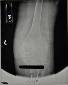
Patient 358/1946 was admitted to Stannington Sanatorium in October 1946 with tuberculous arthritis of the left knee. The patient notes detail that on admittance there was radiographic evidence of destructive lesions already identifiable, however, the first radiographs taken of the individual are of poor exposure or whilst the individual was in plaster cast, so identification is challenging.

The radiographs from February 1947 show the bony anomalies to the knee joint clearly. There is a significant reduction in joint space between the femur and the tibia. The distal epiphysis of the femur shows severe displacement, having moved towards the posterior. Similar displacement can be seen on the proximal tibia to a slightly lesser degree. The patient notes at this stage indicate no change from time of admittance that two sinuses were present above the patella and that immobilisation of the knee was to continue.
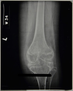
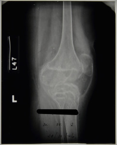
 

HOSP-STAN-07-01-02-1344_05
HOSP-STAN-07-01-02-1344_06


In August 1947, an examination by the visiting physician describes: 

‘Complete disorganisation of the joint. Less decalcification and bony trabeculae are beginning to show.

Fusion of the joint is not complete and there is still some heat.

To be put in plaster for three months’

Changes in the radiographic images between February 1947 and June 1948, when the patient is discharged, are minimal. In December 1947 the physician stated in the patient’s notes:

‘No change in appearance.

There is not complete bony ankyloses of the knee but movement is negligible.

A sinus on the front of the knee which is covered by a scab, is not at present discharging’

There is little or no heat in the knee.

For Thomas’ walking knee splint, patton and crutches.’

No further changes were noted at this stage with the radiographic image below, dated to December 1947, revealing gross anatomical destruction of the knee joint to have taken place and there is no remaining joint space. The striation pattern across the epiphysis and metaphysis of both the femur and tibia is likely to be the result of cartilage destruction and bone degeneration causing porosity in the bones.
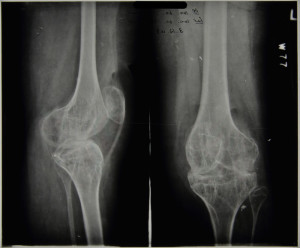
 

HOSP-STAN-07-01-02-1344_13

Patient 358/1946 was discharged in June 1948 but according to their patient notes returned twice as an out-patient and was seen a further two times at the Sanderson Orthopaedic Hospital, Gosforth.


For a case study on the surgical interventions used in tuberculosis of the hip, see earlier post of 08/12/2014

Further radiographic images can be seen on Flickr at https://www.flickr.com/photos/99322319@N07/sets/72157648833066476/


Sources

Albuquerque-Jonathan, G (2006). Atypical tuberculosis of the knee joint. South African Journal of Radiology p.28.

Arthanari, S; Yusuf, S and Nisar, M (2008). Tuberculosis of the Knee Complicating Seronegative Arthritis. Journal of Rheumatology: http://www.jrheum.com/subscribers/08/06/1227.html

Matron’s Medical Report Book – Part 3

Following on from our post on the 12th January we have the third entry from the Matron’s Medical Report Book, with the arrival of another 12 patients and the increase in numbers now beginning to put a strain on the sanatorium’s resources.  By the end of 1908 a total of 62 patients had been admitted and the sanatorium grew considerably over the coming years with the addition of new wings and more beds.

June 12th 1908

“Twelve new patients have come during the last month.

11. Clementine Logan, aged 9; 94, Adelaide St, South Shields

12. Isabella Clementson, aged 12; 123, Robinson St, South Shields

13. George Regan, aged 14; 6, Kyle St, Newcastle-upon-Tyne

14. Hannah E. Hindmarsh, aged 11; 19 Scotch Arms Yard, Morpeth

15. Peter Miller, aged 15; 124, Newgate St, Morpeth

16. Arthur B. Jackman, aged 7 ½; Hanover Sq, Newcastle-upon-Tyne

17. Amelia Seitz, aged 12; 11 Market Place, South Shields

18. Joseph Toward, aged 16 ¼; 9 Carlisle St, Newcastle-upon-Tyne

19. Clara Wilson, aged 11 ½; 12, Newcombe St, Newcastle-upon-Tyne

20. Mary G. Benson, aged 12 ½; 9, Annie Jane Terrace, Gateshead

21. John Gray, aged 15; 12 Pearson St, High Walker

22. Jane A. Farrow, aged 7 ¾; 83, Violet Street, Benwell, Newcastle


Of the four patients whose time is up today application for a further extension has been made in the case of two, Margaret J. Smith & James Robson.  It is hoped that a third J. E. Kenney, may go to the Philipson Farm Colony where the final arrest of his disease might be established.  The fourth, T. Hill, goes home practically ‘cured’.  These last two have gained 6 ½ lbs & 6 lbs respectively in weight.

The general condition of the patients is quite satisfactory.  Most of them are gaining weight rapidly.  Several of the new patients are feverish.  Four have not coughed up any phlegm.  Tubercle Bacilli were present in six of the remaining eight cases.

The children (who are fit to) now do work for about 2 hrs every day & Mr Atkin has kindly prepared a strip of land where they do some light gardening.  We are expecting a private patient in about a week’s time.  Another nurse will then be essential.  An average of 5 to 6 patients in bed (on account of fever) adds considerably to the labour for the nurses & the strain of constantly holding the others in check makes their work very tiring.”


Patients and Staff Outside the Sanatorium c.1920s [HOSP/STAN/11/1/54]

Surgical Procedures – Tenotomy

This week we’re going to take a look at one of the surgical procedures that was used to treat TB and to counter some of it’s side-effects.   A register of operations held within the collection lists a number of different procedures that were carried out on the patients, the majority having been performed by the visiting surgeon Mr Johnston.  These procedures were employed predominantly before the advent of effective antibiotics but many of them still continued to be used effectively after the introduction of drug therapies.  Over the coming months we will explore a range of different treatments that were employed at Stannington, beginning this week with tenotomy.


The records of Stannington Sanatorium show tenotomy to have been performed on several patients suffering from tuberculosis of the bones and joints.  The procedure involved the division of the tendon to lengthen it and would be performed in order to counter deformity caused by the tuberculous disease and increase flexion in a particular joint.  The tendon would be divided, with the two sections allowed to pull apart and then allowed to re-heal at the overlap of the two parts thus resulting in a lengthening of the tendon.  This is not a procedure exclusive to the treatment of tuberculosis but rather in tackling any disease or deformity that has adversely affected the joint.  Patients could find themselves unable to fully flex a particular joint and may be left holding it at an unusual angle owing to muscle contracture and possible subluxation, whereby one muscle pulls more strongly than its counterpart causing partial or complete dislocation of the joint.  Following the procedure the affected joint may be put in plaster or splinted to ensure that the tendon heals at the new length and the joint is in the correct position.


Patient 83/39 was one patient that underwent the procedure, in this particular case division of the hamstrings to tackle poor flexion in the right knee.  Female and aged 3, patient 83/39 was admitted to the sanatorium on 22 October 1937 with a positive mantoux test and the affected knee described as hot, swollen and having limited flexion.  Her pre-admission report reads:

“Fell and after 14 days R knee began to swell and began to limp.  Went to Durham County Hosp. & leg was immobilised in plaster 4 times.  Typical tub. R knee joint probably synovial type.”


Figure 1 - HOSP/STAN/7/1/2/206_01
Figure 1 – HOSP/STAN/7/1/2/206_01


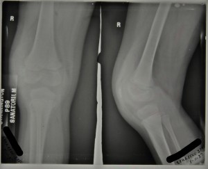
The above radiograph was taken in November 1937, two weeks after admission, with the x-ray report as follows ‘Thickening of capsule.  Eburnation and erosion of femoral condyles, especially outer.’


Her progress is noted in her file throughout her stay and in May 1938 it is reported that the flexion in her knee is at 45 degrees and in August 1938 it is decided that she should have her hamstrings divided with the procedure carried out by Mr Johnston on 17 August 1938.  There appears to be some initial progress as in September 1938 flexion in the right knee is now reported to be 50 degrees and by October she was wearing a Thomas splint (a traction splint commonly used to immobilise the leg).


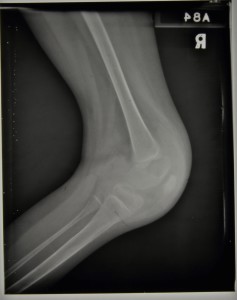
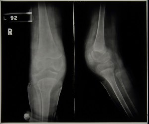
During 1939 progress appeared to be limited with flexion remaining at around 45 degrees throughout, but by January 1940 we see reports that the knee has improved and is almost straight.  In April 1940 she was walking well and she was eventually discharged on 9th August 1940 able to walk in a splint.  It is debateable as to how much impact the division of the hamstrings had on the eventual straightening of the knee and how much was down to the long term splinting and immobilisation of the joint but she was nonetheless eventually discharged as quiescent.  The two radiographs below were taken later on in her stay and date from c.1939/40,


HOSP-STAN-07-01-02-206_04
Figure 2 – HOSP/STAN/7/1/2/206_04
HOSP-STAN-07-01-02-206_02
Figure 3 – HOSP/STAN/7/1/2/206_02


Sources:

J. Krol, Surgery for Deformities Due to Poliomyelitis, (Geneva: World Health Organisation, 1993)