The Stannington Radiographs

The radiographs make up a significant part of the Stannington collection with a total of 14,674 separate images relating to 2220 different patients covering roughly a 20 year period from 1936 to c.1955. When the records were recovered in the 1980s the vast majority of the radiographs were copied on to microfiche and the originals destroyed as they were unstable. However, we still have 326 original radiographs within the collection. Over the course of the project all the microfiche images and the originals will be digitised and made publicly accessible. We also hope to preserve the remaining original radiographs as examples of how x-ray images at the time were produced. The problem here lies with the unstable nature of the film and its natural degradation.


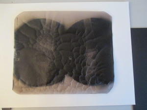
All the radiographs were produced on cellulose acetate film, known as safety film as it replaced the earlier nitrate film that was highly flammable and potentially self-combustible, a problem for many film archives today. Over time the cellulose acetate film naturally breaks down, the early stages of this are recognisable by the strong smell of vinegar coming off the film as the process gives off acetic acid and because of this is known as vinegar syndrome. Eventually as the base of the film and the top layer pull away from one another the film will begin to buckle and crack and bubbles can form under the surface.

IMG_0904
Figure 1
IMG_0905
Figure 2

This process is already evident in several of the radiographs we hold (figures 1 & 2) and unfortunately there isn’t anything that can be done to reverse or halt the process. By storing the films in a closely monitored temperature and humidity controlled environment we hope to delay the process in most of the radiographs for as long as possible.

 

IMG_0903
Figure 4
IMG_0902
Figure 3


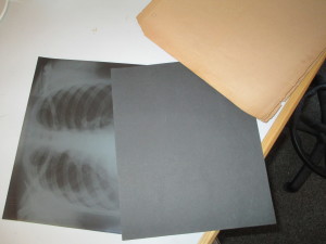
The x-rays were originally stored in ordinary brown envelopes and there could be as many as 15 in each envelope. (Figure 3)  To help preserve them we have instead transferred each x-ray into an individual acid free sleeve. (Figure 4)  By storing them individually we are able to minimise any accumulation of acetic acid that is produced in the degradation process.  Thanks to advice from conservators at Durham County Record Office and their assistance in sourcing the new x-ray envelopes, all the original films are now safely stored in their own envelopes in our photogrpahic strong room.


This degradation is evident in some of the microfiche as well as the original films had obviously started to buckle already at the time they were transferred to microfiche in the 1980s. Consequently some of the images are obscured by a crackling effect. Nevertheless the vast majority of the 14,674 images remain easily readable and the digitisation process will mean that they remain clearly accessible for future use.


[See our Flickr stream for examples of some of the radiographs https://www.flickr.com/photos/99322319@N07/sets/72157648833066476/]

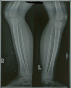
Harris Lines of Arrested Growth

The poor living conditions that many of the children at Stannington Sanatorium came from, outlined in our last post, can often leave physical markers on the skeleton, namely Harris Lines.

Harris Lines are an indication of periods of arrested growth whilst the body is still growing during childhood and can be displayed as opaque, transverse lines on long bones. These can be identified through radiographic imaging or physically on skeletal remains.

The appearance of these lines is considered to show periods in an individual’s childhood when the body comes under stress, which is usually attributed to malnutrition or significant childhood disease. In order for the individual to acquire Harris Lines, they have to have recovered from the period of stress, prolonged malnutrition or disease would not result in their appearance.

HOSP-STAN-07-01-02-1625-06

Numerous patients from Stannington Sanatorium demonstrate Harris Lines in their radiographs. One such example is seen above, patient 148/1948. Thi image was taken in November 1948, approximately 10 months after initial diagnosis of tuberculosis was made. On this patient Harris Lines are identifiable on both proximal and, to a greater degree, distal tibiae, as a brighter, opaque line close to the epiphysis.

This individual was admitted to the sanatorium aged 2 with a Primary Complex, primary infection in the mid zone of the right lung. Their medical history indicates that their father had been diagnosed with pulmonary tuberculosis and subsequently died. The family, consisting of mother, father and two children were living in one room for the majority of the individual’s life, only moving to a two roomed house around the time of their admission to Stannington and on admittance to the sanatorium the child was described in the medical notes as being ‘thin’ and ‘ pale’.

Social conditions such as these would have attributed to the premise of the child undergoing one or more periods of stress during growth and alongside the description of the individual being ‘thin’, malnutrition is possible. A poor appetite or anorexia is often noted in the medical notes of patients as being symptomatic of tuberculosis, again suggesting possible malnutrition. However, the effects of the tuberculosis infection alone would have put the body under due stress and may, therefore, have contributed to the presence of Harris Lines. Both malnutrition and acute tuberculous infection are potential causes of the Harris Lines, alone and in conjunction with one another, and demonstrate the secondary effects that disease and social conditions can have on an individual’s body.

This patient was removed against medical advice approximately one month after admittance, only to be re-admitted seven months later with tuberculosis of the cervical vertebrae. They were eventually discharged in December 1950, two years later, but continued to be seen as an out-patient. No further treatment was required at Stannington Sanatorium.


For more radiographic images, view the ‘Radiographs from Stannington’ on Flickr https://www.flickr.com/photos/99322319@N07/sets/72157648833066476/

Sources

Roberts, C and Manchester, K (2006). Archaeology of Disease (3rd Edition). Cornell University Press.

Social Conditions

Many of the children admitted to the sanatorium came from impoverished backgrounds and had poor living conditions, nearly certain to be a contributing factor to them contracting TB.  Some of the common risk factors for contracting tuberculosis include overcrowding, malnourishment, a weakened immune system, being either very young or very old and a lack of access to medical care to ensure treatment and prevention.  Consequently, the following account from the records of Stannington Sanatorium hardly comes as a surprise and is by no means unusual.


This particular case from the 1940s perfectly illustrates the challenging conditions and the effect of childhood diseases.  The girl, patient 145/1946, was admitted to Stannignton Sanatorium in October of 1945 at the age of 3.  Her case notes indicate that she had already had measles and pneumonia and had been suffering from her present condition of tuberculosis of the right knee for the past 18 months.  After 4 years of treatment she was considered fit for discharge at which point the living conditions she had left behind at the age of 3 are made clear.  The local medical officer reports that

“The home conditions in this case are appalling.  The housing accommodation is only two rooms, in which are already living four adults and five children.”

With this borne in mind the medical staff, unsurprisingly, consider it counterproductive to discharge the girl home and within 3 months she is instead discharged to the Briarmede Nursery in Gateshead.  Medical staff were obliged to take into consideration the living conditions of all patients and consult with local medical officers in the relevant districts before discharging their patients or risk undoing much of the good that had been achieved during the child’s stay in the sanatorium.  Where conditions were not satisfactory children could be discharged to other institutions, as above, or could find themselves staying at the sanatorium longer than was medically necessary, a situation which the doctors were obviously keen to avoid.


For many children coming from backgrounds such as this, being removed to the sanatorium, whilst it may have been difficult being separated from family at such a young age, may in fact have been a blessing in disguise.  Even without the effective drug treatments we have today, the instant improvement in living conditions would have made untold differences to their health and wellbeing.  In each child’s case file it is quite common to see descriptions of their living conditions in their general and family history, taking into account the type of house they were living in, the number of occupants, and the sanitation available.  This information alongside correspondence from the children’s parents requesting support for applications for improved housing gives us a great insight into some of the social conditions across the North East during this period.


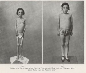
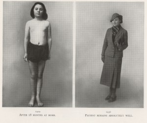
The image below is taken from a brochure produced by the sanatorium in 1936 to promote their services and show images of one patient at four different points in time to illustrate the success of the treatment that Stannington provided. (HOSP/STAN/9/1/1)

NRO 3000-69 PAGE 9 1NRO 3000-69 PAGE 9 2

See also our later post on Harris Lines.