Surgical Procedures – Curettage & Skin Graft

The second in our series of posts on some of the surgical procedures carried out at Stannington focuses on the use of curettage and a skin graft to treat tuberculous skin infections.


Patient 84/37 was male and aged 13 ½ when he was admitted to Stannington on 16th December 1938 diagnosed with TB of other organs and an old ankylosed ankle joint.  He had previously been in the sanatorium from June 1936 to July 1938 suffering from TB of the right ankle which had healed but since his discharge in July 1938 he had developed a tuberculous skin infection on his right ankle overlaying the original tuberculous focus.  This sort of infection might be referred to today as scrofuloderma where there is a direct extension of the tuberculous disease from underlying structures, such as the bone, to the skin.  A report on his condition on admittance reads as follows:


Large sinus R ankle, healed, but skin lower part reddened & thin & scabbed.  Healed sinus R knee & 3 healed on thigh and 1 on leg.  Mobility good’
 

HOSP-STAN-07-01-02-296_07
Figure 2 – HOSP/STAN/7/1/2/296_07
HOSP-STAN-07-01-02-296_04
Figure 1 – HOSP/STAN/7/1/2/296_04


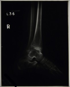
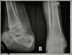
Radiographs taken of his right ankle during his second stay in the sanatorium show the tuberculous ankle to be healed and therefore not causing medical staff any great concern.  Figure 1 is a radiograph taken in1939 for which the report reads, ‘no bone lesion in the right foot’, and figure 2 was taken in 1940 with the report stating that there are ‘bony ankyloses of ankle joint’.


Throughout his stay comments in his case file reveal the scar on his ankle to be thin, unsound and broken down.  Given that at this time there were no antibiotics available to treat this skin infection a commonly used minor surgical procedure was opted for.  On 9th August 1940 curettage was performed on an area on the lateral side of the right ankle with a Thiersch skin graft.  Curettage simply refers to the removal of the infected tissue using a surgical tool called a curette.  A Thiersch skin graft is a split-thickness graft that can be quite thin and involves the removal of the epidermis and part of the dermis from a donor site elsewhere on the patient’s body, which can then be placed in narrow strips over the wound.  By November of 1940 it was noted that the skin graft had taken well, was soundly healed, and that there was good movement of the foot at the 1st metatarsal joint.  He was discharged quiescent on 19th November 1940 with the procedure having been a success.


Sources:

B. Kumar and S. Dogra, ‘Cutaneous Tuberculosis’, in Skin Infecitons: Diagnoisis and Treatment, Edited by J. C. Hall and B. J. Hall (Cambridge: Cambridge University Press, 2009)

L. Teot, P. E. Banwell, & U. E. Ziegler, Surgery in Wounds, (Berlin: Springer-Verlag, 2004)

Safe Milk Supplies

We touched upon the problems of infected milk supplies in a previous posting on abdominal TB and we’ll focus on the issue in more detail here.  Mycobacterium bovis is the pathogen responsible for the development of TB in cattle, which is commonly referred to as bovine TB.  The consumption of milk from cows infected with bovine TB and in turn the ingestion of mycobacterium bovis can lead to an individual developing TB.  This was for many years a very common cause of TB in humans and remains so in countries that do not routinely pasteurise milk.  Pasteurisation involves the heating of the milk in such a way as to kill off any bacteria that might be present, and through its use the spread of bovine TB to humans has nearly been eradicated in the UK.


The 1875 Public Health Act made it compulsory for local authorities to appoint a Medical Officer of Health (MoH) who produced an annual report detailing any health and sanitary issues in the district as well as giving a wealth of statistical information related to birth and death rates, population, infectious diseases and causes of death.  The MoH for Northumberland makes regular reports on the situation in the county regarding tuberculosis including comments on the causes of abdominal tuberculosis and efforts made to prevent its spread.  In the 1906 report he states:

“That about 30 per cent of the milch cows in England are tuberculous, and that consequently infants and persons suffering dangerous illness are in many cases being fed milk containing the organisms of tuberculosis” [NRO 3897/3, 1906 p.21]

The problem of infected animal products is clearly recognised by medical and sanitary officials early on in the 20th century but little is done to tackle the situation head on and so abdominal tuberculosis continues to be a significant problem.  Three years later in 1909 the MoH expresses his frustration at the situation and lack of power to change it:

“The elimination of tuberculosis from dairy herds is a matter of great difficulty since, at present, no assistance is given, by the state, to the farmer who, for the benefit of the general public as well as for his own advantage, may wish to obtain a herd free from this disease.”  [NRO 3897/3, 1909 p.33]

It is not until the 1940s that significant steps were taken to introduce tuberculin tested milk and encourage pasteurization.

“The eradication of tuberculosis from our milk supplies is a matter of greatest importance to us all, and it is encouraging to note the marked increase in the production of milk from tuberculin tested cows.  45% of all the milk produced in the County was from such herds, and it is known that in 1948 the proportion had risen to more than 50%.” [NRO 4081/1, 1947 p.8]


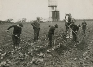
HOSP/STAN/11/1/51 Boys at work on the farm
HOSP/STAN/11/1/51
Boys at work on the farm


Milk supplies were something given great consideration by those responsible for the establishment of Stannington Sanatorium from the outset.  In 1905, two years before the official opening of the sanatorium, a farm colony was established on the site to take in young boys and provide them with training.  It was from here that the sanatorium was able to receive a safe supply of milk from tuberculin tested cows.  Tuberculin testing is another method used in preventing the spread of bovine TB whereby the cows were tested to see whether they carried mycobacterium bovis rather than treating the milk itself.  This method was used quite commonly early on before the onset of widespread pasteurisation and would have been essential to the recovery of many of the patients and in preventing any of them acquiring any further infection.  As time goes on, and tuberculin testing and pasteurisation is implemented more widely across the county, it is notable when looking at the patient files that instances of abdominal TB decrease particularly as we enter the 1950s.


Sources:

ALLISON, T. M. (1908) Children’s Sanatorium, Stannington, Northumberland, British Journal of Tuberculosis, 2 (3), p.204

SCHOFIELD, P. F. (1985) Abdominal Tuberculosis, Gut, 26 (12), pp.1275-1278

NORTHUMBERLAND ARCHIVES: NRO 03897, Northumberland County Council: County Medical Officer of Health Reports, 1893-1935

NORTHUMBERLAND ARCHIVES: NRO 04081, Northumberland Health Authority: Records, 1942-1970

Patient 90/38, An Unconfirmed Diagnosis

Amongst the patients admitted to Stannington Sanatorium there are a number for which following admission doctors decide that their condition for whatever reason is non-tuberculous.  Differential diagnoses can vary from bronchiectasis and asthma in those suspected of having pulmonary TB to Perthes’ Disease in those suspected of having TB of the hip.  One patient who ultimately appears not to have TB is patient 90/38, a 17 ½ year old girl presenting with strong neurological symptoms, although no definite conclusions seem to be drawn on what the cause might be.


Admitted on 12 Sept 1941, she is one of the very few private patients and also one of the oldest.  The diagnosis given at the top of her file is ‘Non-TB, query bone tumour spine and skull’.  She had been suffering from symptoms for a year prior to admission and reports immediately following admission state ‘Lower thoracic curvature, no active angular deformity.  Not tuberculous’.


The first x-rays of her spine are taken the day after admission and here the report reads:

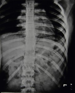
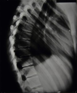
Marked irregularity of epiphyses in lower thoracic region.  Some wedging of bodies of 9th and 10th dorsal vertebrae.  Edges of bone are irregular & ossification is either incomplete or of poor quality.

                Diagnosis: Epiphysitis of thoracic region, probably not tubercular

Over the coming months further spinal x-rays and their corresponding reports do not suggest any significant worsening of the spinal wedging nor any great improvements.  The final report indicates that 5 vertebrae are affected with the 9th and 10th being the worst.  Figures 1 and 2 are examples of some of the spinal x-rays that were taken.


HOSP/STAN/7/1/2/651_25
Figure 1 – HOSP/STAN/7/1/2/651_25
HOSP/STAN/7/1/2/651_03
Figure 2 – HOSP/STAN/7/1/2/651_03


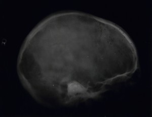
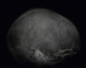
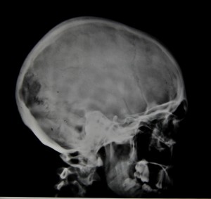
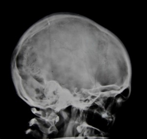
In addition, x-rays were taken of her arms, forearms, pelvis, femora, and legs, all of which were clear.  There are also 7 x-rays taken of the skull, 4 of which can be seen in figures 3-6. Reports on the skull x-rays read as follows:

9/12/1941: Skull, localised deficiency of internal table to left of midline – lying over leg area.

19/3/1942: Outline of internal table broken for about 1” in anterior-parietal region. 

14/5/1942:  Rarefaction appears to be falling in.  Outline more normal.  Break still about 1”. 

Figure 3 - HOSP/STAN/7/1/2/651_23
Figure 3 – HOSP/STAN/7/1/2/651_23
Figure 4 - HOSP/STAN/7/1/2/651_14
Figure 4 – HOSP/STAN/7/1/2/651_14


Figure 5 - HOSP/STAN/7/1/2/651_21
Figure 5 – HOSP/STAN/7/1/2/651_21


Figure 6 - HOSP/STAN/7/1/2/651_04
Figure 6 – HOSP/STAN/7/1/2/651_04


Her file also contains quite detailed reports on other tests carried out and her general condition during her stay.  In November 1941 reports are made of signs of mental disturbance and that she ‘will not speak to anyone and only laughs or cries when spoken to’.  She is also experiencing some incontinence and has a history of incontinence between the ages of 8 and 14.  She has bilateral ankle clonus and a positive Babinski test, more marked on the right.  Two days later the report reads as follows:

Spasticity lower limbs.  KJs +.  Bilateral ankle clonus.  Plantar Reflex? – probably flexor.  Sensation apparently normal.  Pupils reacting normally.  Eye movements, other cranial nerves & field of vision-apparently normal but patient unresponsive & difficult to examine.

She says she feels miserable & that everyone thinks she is silly, & that she has been like this before.

Still some incontinence.


At the end of November 1941 it is noted that there is a white patch in the centre of the optic discs and that the disc edges are blurred, still some spasticity, slight clonus, sluggish Babinski, normal co-ordination, normal mental condition, and occasionally experiences some frontal headaches.  In March of 1942 a Wassermann Test comes back negative and she is eventually discharged on 16th May 1942.

If anyone can offer any further opinions on the possible causes of her condition please feel free to add your comments below.