🎄Christmas Opening Hours for 2024🎄
CLOSED between 4pm Friday 20th December 2024 and 10am Thusday January 2nd 2025
Ordinary opening hours apply before and after this period.

Streptomycin

Selman Waksman New York World-Telegram and the Sun staff photographer: Higgins, Roger, photographer/ Wikimedia Commons/ Public Domain
Selman Waksman
New York World-Telegram and the Sun staff photographer: Higgins, Roger, photographer/ Wikimedia Commons/ Public Domain


Streptomycin was the first antibiotic drug to be discovered that was effective in the treatment of tuberculosis.  It was isolated in October 1943 by Albert Schatz, Selman Waksman, and Elizabeth Bugie  with Waksman going on to win the Nobel Prize for Medicine in 1952 for his work on the discovery of streptomycin.  Extensive human trials of the drug were carried out in the USA in the years following its discovery and the UK’s Medical Research Council (MRC) carried out its first randomised, controlled clinical trial of the drug in 1946.  The MRC’s trial aimed to compare the effectiveness of streptomycin combined with bed rest with that of bed rest alone and did eventually show the drug to be more effective.


At this point the drug was used in conjunction with the traditional methods utilised in the sanatoriums, such as bed rest and light treatment, and we start to see cases of streptomycin being used as treatment in Stannington Sanatorium from 1947.  Although it was available as an effective treatment and the only drug treatment option it was not widely used on the children of Stannington, and instead particular cases were singled out as suitable candidates for treatment.  There were several problems arising from the use of streptomycin that meant it could not be a cure-all treatment for everyone.


The drug must be administered by injection which could prove to be very painful, a particular problem where children were involved.  One girl, patient no. 13/1949, had been receiving regular streptomycin treatment at Newcastle General Hospital before being admitted to Stannington.  Initially intramuscular and intrathecal treatment was used, which involved administering the drug directly into the muscle and into the membrane of the spinal cord.  Daily treatments were continued for 4 weeks and although there were some initial signs of improvement toward the end of the 4 weeks the patient began to become very ill with continuous vomiting, drowsiness, incontinence and papilloedema (swelling of the optic discs caused by intracranial pressure) so treatment had to be stopped.  A week after treatment was stopped there was a marked improvement in her general condition and so treatment was resumed with a general anaesthetic being required for each intrathecal injection.  The patient continued to improve but the papilloedema persisted and the intrathecal therapy was proving difficult to administer.  Instead a tube was inserted along the floor of the skull to the interpeduncular fossa and streptomycin injected on alternate days, which in turn led to the reduction of the papilloedema and improvement in her condition generally.  She was continued on intramuscular injections up to her discharge to Stannington Sanatorium where she was to receive more traditional treatment and rest on the basis that she would be returned to NGH if any relapse in her condition was experienced.


This case clearly illustrates how streptomycin was not a simple cure not least because the administration of the drug was particularly uncomfortable but also because of the side-effects that could be experienced.  One noted side-effect in children is the possibility of irreversible auditory nerve damage.  Contemporary studies also showed that toxic reactions to interthecal streptomycin could occur sometimes with fatal consequences.  The invasive methods of administering the drug meant that when it was first introduced some of the children in Stannington Sanatorium that were chosen to receive the treatment had to be discharged to a local hospital to receive it.  Nonetheless, it still provided incredibly successful results and patient 13/1949 went on to be discharged as quiescent.


Of the cases from Stannington Sanatorium that received streptomycin treatment we can see that they were all suffering from quite severe forms of tuberculosis making streptomycin a last attempt where it was known that traditional sanatorium methods would not work.  For example, the above case, patient 13/1949, was suffering from TB meningitis, which along with miliary TB was responsible for a large number of deaths.   Looking at patient files from the beginning of the 1940s we can see that it was these sorts of cases where deaths regularly occurred, whereas most other manifestations of TB responded well to sanatorium treatment.  In this respect streptomycin was incredibly successful in treating patients that only a couple of years earlier would most likely have died.


The years following the introduction of streptomycin saw the development of several other drugs effective in the treatment in TB which helped to tackle problems of drug resistance that had been developing.  Instead combination therapy using multiple drugs became possible and their proper administration meant that the development of drug-resistant strains could be tackled.  Owing to drug resistance and its difficult administration streptomycin is no longer a first line drug but remains on the World Health Organisation’s (WHO) list of essential medicines.


Sources:

SCHATZ, A, BUGIE, E, & WAKSMAN, S. A. (1944) Streptomycin, a substance exhibiting antibiotic activity against gram-positive and gram-negative bacteria, Proceedings of the Society for Experimental Biology and Medicine, 55, pp.66-69.

BYNUM, H. (2012) Spitting Blood: The History of Tuberculosis, Oxford University Press, p.195.

MILLER, F. J. W, SEAL, R. M. E, and TAYLOR, M. D. (1963) Tuberculosis in Children, J & A Churchill Ltd. p.184.

Matron’s Medical Report Book-Part 4: A Bumpy Start

Given the mostly glowing reports we’ve heard about life at the sanatorium from past patients from the 1930s & 1940s who recounted their experiences in a recent oral history project it is surprising to learn from some of the earlier written accounts that the sanatorium wasn’t always so perfect.


April 17th 1909

“I found the sanatorium in an unexpected condition, distinctly neglected, especially the kitchen, back kitchens, store cupboards & meat safe.  The food almost unfit to eat, badly cooked & cold.  In the last few weeks things have improved somewhat, especially the food.  The cases in the sanatorium at present would seem to be improving – 9 in bed against 19 a few weeks ago.”


This report is seemingly made by one of the early matrons and her subsequent reports over the next few years begin to tell of satisfactory conditions.  However, following the appointment of a new matron in 1912, the story again seems far from satisfactory.


April 1912

“On Tuesday March 26th I took up the duties of ‘matron’ in the sanatorium.  Miss Linton, the acting matron, left the following morning.  Acting on the advice of Dr Allison, I engaged a temporary sister & advertised for a permanent one.  I secured the services of Nurse Batty from the Granville Nursing Home, who on April 6th accepted the appointment of sister to the sanatorium, at the salary of £40 per year.  Sister has trained at the Royal Victoria Infirmary and is helping me in my endeavours to bring the sanatorium to a recognized hospital standard…

There having been no permanent head in the sanatorium since January, there was some little excuse for the condition of the place.

The nursing staff, the maids and the children were decidedly out of hand.

I have employed two maids to clean the sanatorium & request that I may keep one when the cleaning is finished, as the work in the kitchens & attics cannot be done with the present staff…

As regard the children (there are 59 patients in the sanatorium).  They need to be taught sanatorium methods; as a beginning I have kept all the children in bed for a week until they show signs of gaining weight & of going quietly in everyway.”

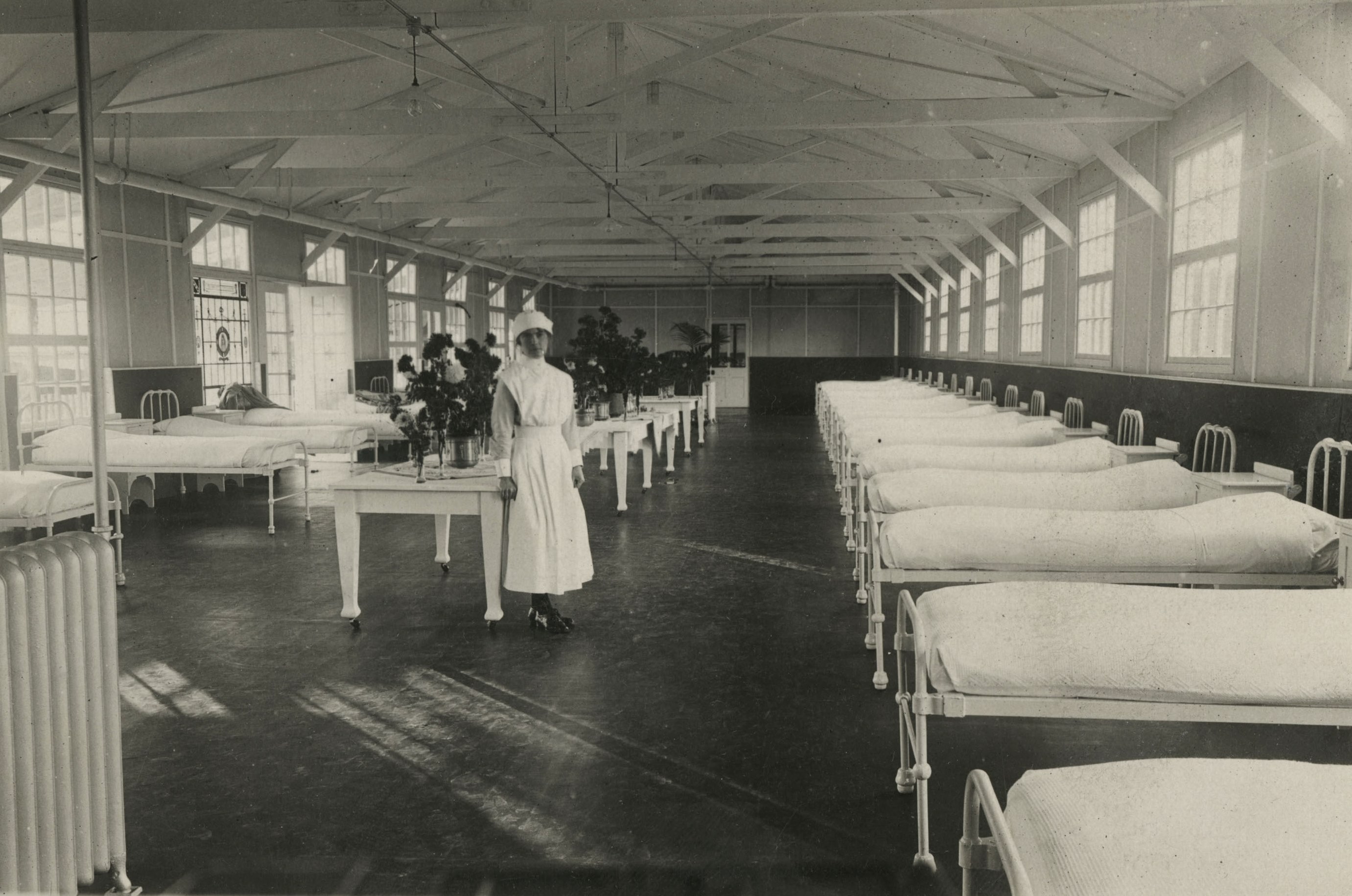
A nurse on one of the wards, HOSP/STAN/11/1/55


The new matron, S.M. Robson, continued to work at the sanatorium until 1914, during which time it seems that there were continued efforts to improve the conditions and the operation of the sanatorium.  The following years saw a great many changes made to the sanatorium with several charitable donations leading to further extensions and a huge increase in the sanatorium’s capacity. With the expansion came the addition of new facilities such as the vita glass pavilion and the x-ray department.  It seems that this along with the concerted efforts of the new matron meant that the sanatorium was able to grow into a successful and well-functioning hospital.

Osteomyelitis Part 2: Dactylitis

Continuing on from our last post on osteomyelitis affecting the lower leg bones, see post dating 06/02/2015, here we are going to review a case of tuberculous osteomyelitis of the short tubular bones in the hands and feet; the metacarpals, metatarsals and phalanges,  commonly known as tuberculous dactylitis or ‘spina ventosa’(meaning short or small bone inflated with air). This is a particularly uncommon manifestation of tuberculosis primarily affecting children, and it is rare in anyone over the age of six.

Dactylitis affects the hands more often than the feet and can affect multiple bones at one time. It is caused by the haematogenous spread of tubercular bacteria which settles in the bone marrow of the short bones prior to the epiphyseal centre becoming established. This leads to thickening of the periosteum (outer membrane of the bone) with osteomyelitis, but rarely involves the joint.


Patient 90/27

This patient was a 16 year old male, admitted to Stannington Sanatorium in September 1940 with tuberculosis of the bones and joints, stage II. In this instance tuberculous dactylitis was diagnosed affecting the left foot and right hand, alongside queried primary infection in the lungs and concerns over the right elbow.

The patient’s medical history states that seven months prior to his admission the patient’s left ankle became swollen and started discharging; his 4th left toe became swollen and started discharging and 1 year prior to admission his right hand was hurt and it too became swollen.

Initial observations made by admitting doctors read as follows:

‘Left foot sinus over lateral malleolus,

swelling over 4th toe left foot, discharging sinus at  base,

right hand hard swelling of 5th metacarpal’


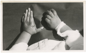
Diagnosis of dactylitis is made based on radiographic findings; however, it is often observable physically due to painless inflammation of the soft tissue surrounding the affected bone. As noted above sinuses may also form, which may discharge, as a result of infection. Although we have no photographic images of patient 90/27, we do have a photograph of another patient (for whom we have no radiographs) also diagnosed with tuberculous dactylitis showing the effects this infection had on the surrounding soft tissue, note the presence of a discharging sinus at the base of the first finger on the left hand, Figure 1.

FIGURE 1: HOSP-STAN-07-01-01-361_06


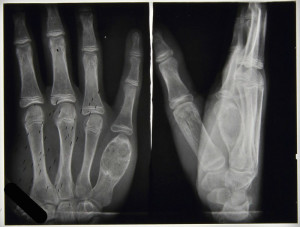
The first x-ray report for patient 90/27 was in October 1940 and confirmed that the phalange of the fourth toe of the left foot was expanded but without any signs of a cavity; the fibula showed signed of decalcification; fibrosis was detected in the lungs, possibly the primary source of the tubercular infection, and the fifth metacarpal of the right hand was badly affected, Figure 2.


FIGURE 2: HOSP-STAN-07-01-02-641_07

Once established, the tuberculous infection quickly involves the entire marrow space and the tuberculous granulation tissue expands the bone cortex following necrosis of the bone tissue. As a result the bone expands taking on a spindle form and appears much like an inflated balloon. This is well demonstrated in Figure 2, with the balloon like inflammation in the distal metacarpal. It is common to see new bone formation, or periostitis, as a result of the infection. Soft Tissue swelling can also be seen surrounded the affected metacarpal in Figure 2.


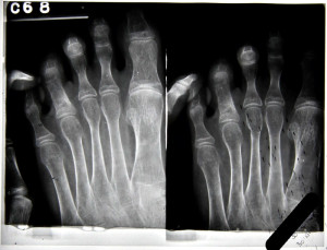
FIGURE 3: HOSP-STAN-07-01-02-641_11

Throughout the patient’s notes, specific areas of infection are focussed upon. In April 1941 the x-ray report notes look at the fourth toe of the left foot, Figure 3. Here the proximal phalanx is noticeably expanded and the notes state that the cavity looks as though it has been filled in with granular tissue. By February 1942 the disease has taken over the whole of the phalanx and a cavity is noted in the distal end of the bone.

There is nothing within the patient notes about any specific treatment this patient was receiving for his condition. Given the nature of the infection and the continuous references to ulcers and sinuses that were discharging it is likely these would have been drained regularly as part of the general sanatorium treatment, alongside rest and fresh air. There is one side note within the notes that questions excision of toe, however this is not pursued anywhere else.

FIGURE 4: HOSP-STAN-07-01-02-641_05


With tuberculous dactylitis, it is possible to achieve almost complete recovery. New bone formation around the affected bone is noted, but soft tissue swelling abates and deformity is rare, Figure 4. In April 1942 this patient’s notes read:

‘Nil active in lungs.

Foot: cavity in bone of 4th phalanx filled up. Quiescent.

Hand: metacarpal improving’


This patient was later discharged in May 1942 as ‘improved.’


Further radiographic images can be seen on the Stannington Sanatorium ‘Radiographs from Stannington’ Flickr stream https://www.flickr.com/photos/99322319@N07/sets/72157648833066476/


Sources

Bhaskar, Khongla, T and Bareh, J (2013). Tuberculous dactylitis (spina ventosa) with concomitant ipsilateral axillary scrofuloderma in an immunocompetent child: A rare presentation of skeletal tuberculosis. Advanced Biomedical Research 2:29

Mishra Gyanshankar, P, Dhamgaye, T.M.  and Fuladi Amol, B (2009). Spina VentosaDischarging Tubercle Bacilli – A Case Report. Indian Journal of Tuberculosis 56: 100-103

Roberts, C and Buikstra, J (2003). The Bioarchaeology of Tuberculosis: A Global View on Reemerging Disease. Univesity Press of Florida.