🎄Christmas Opening Hours for 2024🎄
CLOSED between 4pm Friday 20th December 2024 and 10am Thusday January 2nd 2025
Ordinary opening hours apply before and after this period.

Case Study – Tuberculosis of the Hip

The radiographs from the Stannington collection allow for a detailed insight into the effects that tuberculosis had on the body. One such example is patient 132/1951, a nine year old individual transferred from Fleming Memorial Hospital to the Stannington Sanatorium with Tuberculosis of the Hip. There are a total of 31 radiographic images allotted to this individual, mostly of the pelvic area but also some of the right knee.
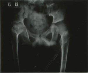
 

HOSP-STAN-7-1-2-2046-16
Figure 1 – HOSP/STAN/7/1/2/2046


Infection is evident in the right hip, the femoral head (head of the femur) and the acetabulum (socket of the pelvis) show visible signs of bone destruction which continues down into the lower part of the pelvis, the ischium. The reduced gap in the joint between the femoral head and the acetabulum is also indicative of tuberculosis (Figure 1). There is also some porosity shown in the femoral head, which displays the weakened state of the bone due to the extent of the infection.

This can be compared to the healthy, left side of the pelvis where a clear ball shaped femoral head can be distinguished with structured shape, lined up with the acetabulum. The gap between the upper and lower sections of the pelvis is evidence of this being a child as the pelvis has not yet fused.

Due to the level of destruction to the right hip, there would have be a significant impact on this individual’s standard of living, the possibility of reduced mobility due to ankylosis of the hip and atrophy.
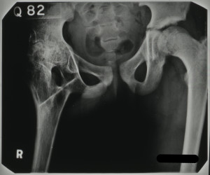
NRO-3000-HOSP-STAN-07-01-02-2046-30
Figure 2 – HOSP/STAN/7/1/2/2046


Figure 2, however, shows the results of a surgical intervention to repair damage caused by the tuberculosis infection, a procedure known as arthrodesis. This procedure is descried in the patient’s notes in a letter from the surgeon at the Royal Victoria Infirmary, Newcastle to a doctor at Stannington:

‘… a cortical graft was taken from the anterior aspect of the right tibia. This would was closed with catgut and silk-worm gut to the skin. The right hip was approached from an incision over the posterior aspect of the greater trochanter…. The shaft of the femur was divided just below the greater trochanter and a gap made in the ischium. The bone graft was inserted into two gaps between the femur and the ischium…..’


As a result of the surgery it is noted that there was some flexion in the right hip and some apparent lengthening of the right leg. The tuberculosis infection was deemed quiescent and this individual, after being monitored as an outpatient, went on to be discharged as ‘healed’.

View more radiographs on our Flickr stream https://www.flickr.com/photos/99322319@N07/sets/72157648833066476/

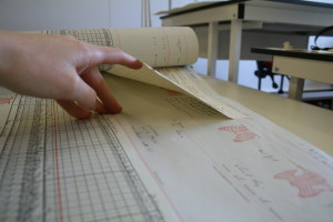
Conservation Work on the Early Patient Files

The earlier patient notes from Stannington Sanatorium were bound together in what were known as ‘Discharge Books’.  We had 15 of these large volumes and took the decision to have them unbound and to keep them stored as individual sets of notes instead.  They still remain in their original order but thanks to the work of the conservators at Durham Record Office are now much more accessible and neatly stored in their own custom-made boxes.  The conservators have very kindly given us a step-by-step account of the work they carried out on these records:


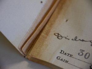
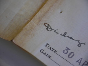
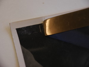
During their time at the sanatorium each patient had annotated medical notes charting their progress.  These came in a standard format; a bifolio (a sheet of paper folded in half to make two leaves), often with additional sheets or photographs attached. After the patients had been discharged these bifolios were bound together into volumes.    Whilst keeping the notes safe, the bindings were very tight, meaning it was difficult to read text written in the fold or ‘gutter’.  Furthermore, small pieces of paper that were adhered to the notes were often trapped within the gutter, unable to be lifted and thus obscuring text beneath them. (Figure 01)

03  'Trapped' paper obscurring text
Figure 01 – ‘Trapped’ paper obscuring text


Whilst normally all care is taken to preserve bindings as part of an object’s historical context, the decision was made that the information that was inaccessible due to the binding structure was more important than the bindings themselves.  As such, I and two Conservators at Durham County Record Office have been dis-binding the volumes.


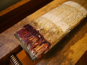
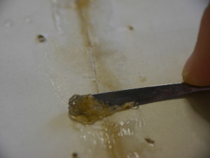
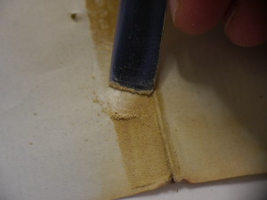
Dis-binding begins with removing the boards and mechanically stripping as much leather from the spine as possible.  The leather was adhered directly to the text-block paper with a thick animal glue.  To remove the glue a poultice of sodium carboxymethyl cellulose was applied which gradually softens the animal glue without wetting the paper beneath.  When the glue was sufficiently softened we gently scraped it away, exposing the sewing and paper beneath.  (Figure 02)

05  Application of sodium carboxymethyl cellulose
Figure 02 – Application of sodium carboxymethyl cellulose


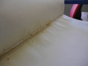
We were then able to cut the sewing and separate one bifolio at a time from the rest of the text-block.  In doing so we were able to see the stab sewing the bookbinders had used, which was the reason it was impossible to open the book properly to access the text in the gutter.  When each bifolio was detached, we removed any remaining animal glue from the fold. (Figures 03 & 04)                                                                                    

06  Stab sewing
Figure 03 – Stab sewing
07  Removal of glue from fold
Figure 04 – Removal of glue from fold


Patients who had spent many years in the sanatorium had additional sheets added to their notes.  These were attached to their bifolios with a linen tape.  Over time the adhesive of the tape has failed, becoming dry and grainy.  Whilst this meant the tape carrier was very easy to remove, the adhesive remained on the paper and had to be manually scraped off each sheet. (Figures 05-07) 

Figure 5
Figure 05 – ‘Before’ linen tape
Figure 6
Figure 06 – Removal of adhesive from tape
Figure 7
Figure 07 – ‘After’ tape removal


Whilst most of the paper is still in very good condition, the bifolios with many additional sheets were found to have split in the binding process as the paper could not stretch over the bulk of the inserts.  Furthermore, the inserts often protruded further than the rest of the text-block and so had suffered damage along the edges.  Some damaged bifolios had been ‘repaired’ with tape or paper, and often these repairs were failing.  When collections are being digitised, it means the originals will be rarely accessed.  As such conservators carry out minimal intervention, or repairs for ‘once-only’ handling.  This means we only repair a tear or damage if text is obscured or if we think a tear might be caught and worsened in the digitisation process.  We carried out these repairs with a thin but strong Japanese paper and wheat starch paste. (Figures 08 & 09)

12  'After' paper repair
Figure 09 – ‘After’ paper repair


11 'Before' repair - with an old repair piece
Figure 08 – ‘Before’ repair – with an old repair piece


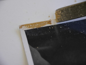
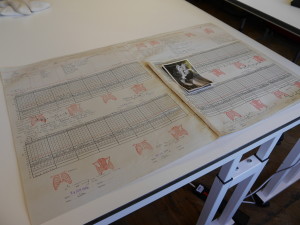
 There were also a number of photographs within the medical notes, adhered with a similar linen tape.  The adhesive was gently removed to prevent scratching the emulsion layer.  Four-flap folders were made out of SilverSafe paper; an unbuffered paper specifically for storing photographs to prevent acidic decay and abrasion. (Figures 10-12)

15  Removal of tape I
Figure 10 – Removal of tape I
16  Removal of tape II
Figure 11 – Removal of tape II
17  'After' page with photographs
Figure 12 – ‘After’ page with photographs


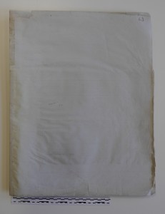
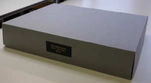
Bespoke clamshell boxes were made from acid-free archival card to house each volume, which are then ready to be digitised. (Figure 13)

19  Archival clamshell box
Figure 13 – Archival clamshell box


Jenny Halling Barnard, Lisa Handke, and Zoe Ross – Conservators at Durham County Record Office

Matron’s Medical Report Book – Part 1

The Matron’s Medical Report Book is the earliest surviving record we have from Stannington Sanatorium, which contains monthly updates on patient progress and general administration.  The first entry from the Report Book, below, dates from 1908 and lists some of the earliest patients with details of their ages, addresses and condition whilst in the Sanatorium.    


April 11th 1908

“There are now six patients here.  Five have been here for nearly four weeks (since March 18th) & one for three weeks.  They are:-

1. Maggie Smith, aged 17.  Address 73 Leopold St (sent from the Gateshead Workhouse)

2. John Edward Kenney, aged 15 ¾; Address 72 William St, Hebburn Quay (working at Hebburn Colliery)

3. James Robson, aged 13. Address 16 Bottle Bank, South Shields

4. Thomas Hill, aged 12 ½. Address 76 Belvedere St, Heaton

5. John Nicholson, aged 10.  Address 10 Hyiamais Court, Gateshead

6. Thomas Burns, aged 16 ¾. Admitted March 21st. Address 40 Stanley St, Jarrow


The general condition of all the patients has considerably improved.  They have all gained in weight during the last 18 days: Maggie Smith 5 ¼ lb, John Kennedy 4lb, James Robson 2 ¾ lb, T. Hill 3lb, J. Nicholson 3 ¾ lb, T. Burns 3 ¼ lb (all given to the nearest ¼ lb).  Only one patient, Maggie Smith, has any marked degree of fever.  There is a slight improvement in her temperature.  One patient, J. Hill, does not cough at all.  All the others are coughing less and bringing up less sputum.

Examination of the sputum for tubercle bacilli has not yet been possible.

The local condition in three patients, T. Burns, T. Hill & J. Nicholson is quite satisfactory, shewing improvement. In two patients, Maggie Smith & J. Robson it has remained about the same.

In one, J. Kennedy, there has been some extension of the diseased area, but improvement again during the last four days after strict rest.”

[HOSP/STAN/2/1/1]


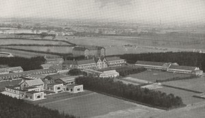
The close association of tuberculosis with poor living conditions is further highlighted here by the first two patients who have come from Gateshead Workhouse and Hebburn Colliery respectively.  At this stage there were only 6 patients but the sanatorium had opened not long before this with provision for 40 children and soon saw the beds fill up.  In the following years the sanatorium’s capacity began to expand rapidly with an extension providing an additional 50 beds opening in 1911 and several new wards opening in the 1920s so that by 1926 there were 312 beds available.

HOSP-STAN-9-1-1 aerial view

Aerial view of the sanatorium c.1936

[HOSP/STAN/9/1/1]

We will post some more entries from the Matron’s Report Book in future blog posts to track the progress of the early days of the Sanatorium.