Mr Henry Mulrea Johnston

Henry Mulrea Johnston was born in County Down, Ireland in 1877 and studied medicine at Queen’s College, Belfast graduating in 1903.  He went on to study and work at Trinity College, Dublin before moving to London in 1910 where he worked at St Bartholomew’s and Great Ormond Street Hospitals and became a fellow of The Royal College of Surgeons in 1911.  In 1912 he was appointed Resident Medical Officer at The Royal Victoria Infirmary in Newcastle.


During WWI he joined the RAMC and was posted to a hospital in Sidcup where he concentrated his efforts on deformities of the face.  Prior to joining the army he had been committed to research using the latest radiological techniques for diagnostic purposes, something that was evident in his research for many years.  An image in an article from the British Dental Journal demonstrates some of the pioneering work that Johnston was involved in whilst at Sidcup and it is also possible that it is Johnston who is pictured on the right of the picture. http://www.nature.com/bdj/journal/v217/n6/full/sj.bdj.2014.820.html
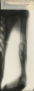
14 year old male with cyst of the humerus treated with with intra-medullary and osteo-periosteal grafts.  6 weeks after operations HOSP/STAN/10/1/30/3
14 year old male with cyst of the humerus treated with with intra-medullary and osteo-periosteal grafts. 6 weeks after operation
HOSP/STAN/10/1/30/3


Following the war he was also appointed Visiting Surgeon to Stannington Children’s Sanatorium, a role which he carried on until 1945.  In his obituary from the British Medical Journal it is remarked that “In general surgery he loved to demonstrate bone tumours and cysts and to illustrate his cases with beautiful radiographs of his own taking.”  Amongst the records of Stannington Sanatorium we have a selection of photographic copes of some of Mr Johnston’s x-rays reflecting some of his interests outside of the Sanatorium.  The collection of images shows patients of varying ages with different skeletal deformities, most of which appear to be unrelated to tuberculosis.
Male, age 8.  Perthe's disease affecting femoral head. HOSP/STAN/10/1/24/1
Male, age 8. Perthes’ disease affecting femoral head.
HOSP/STAN/10/1/24/1
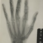
Female, age 22. Right hand. HOSP/STAN/10/1/21/1
Female, age 22. Right hand.
HOSP/STAN/10/1/21/1

Click on images to enlarge


Sources:                                                                                                                                                                           ‘Henry M. Johnston, F.R.C.S’, British Medical Journal, Vol. 2, No. 4724, 21 Jul 1951, pp. 181-182

British Dental Journal http://www.nature.com/bdj/journal/v217/n6/full/sj.bdj.2014.820.html  [24 Nov 2014]

Unreported X-Rays

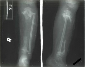
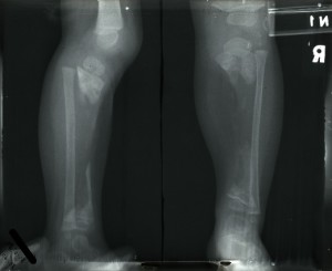
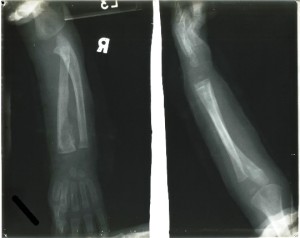
Amongst the 2242 sets of patient radiographs we hold there are 55 for which we do not have any corresponding patient case notes.  Within this group of 55 there is one patient in particular that stands out as the radiographs that survive for him are the earliest original radiographs we hold with the others all being on microfiche.  The other 54 also have their patient numbers clearly marked on the radiographs and references to them can be found amongst the other records in the collection, for example admission registers and treatment registers.


All we know about this patient is his name and the date on which the x-rays were taken.  The three x-rays were taken in September and October of 1939 and show the boy’s right arm and right leg.  Judging from his size we are assuming he was relatively young and was evidently suffering from disease of the right tibia and right forearm.  There are no later re-admissions for this patient and so we are assuming that he either recovered and was discharged with no later relapse or that he died.  We have the sanatorium school records for the period as well and there is no mention of him there, so again we can assume he was either below school age or too ill to attend.  The only other contemporary records we have where we might expect to find him are the register of operations and the register of splints and appliances.  Again there is no reference in either of these confirming he did not receive any surgical treatment during his stay and was not issued with a splint.


HOSP-STAN-7-1-2-2-1 HOSP-STAN-7-1-2-2-2

HOSP-STAN-7-1-2-2-3Click on the images to enlarge


If you can offer any additional details about this patient from his radiographs please add them to the comments below.

Case Study – Tuberculosis of the Hip

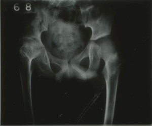
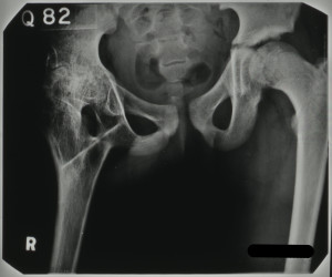
The radiographs from the Stannington collection allow for a detailed insight into the effects that tuberculosis had on the body. One such example is patient 132/1951, a nine year old individual transferred from Fleming Memorial Hospital to the Stannington Sanatorium with Tuberculosis of the Hip. There are a total of 31 radiographic images allotted to this individual, mostly of the pelvic area but also some of the right knee.


HOSP-STAN-7-1-2-2046-16
Figure 1 – HOSP/STAN/7/1/2/2046


Infection is evident in the right hip, the femoral head (head of the femur) and the acetabulum (socket of the pelvis) show visible signs of bone destruction which continues down into the lower part of the pelvis, the ischium. The reduced gap in the joint between the femoral head and the acetabulum is also indicative of tuberculosis (Figure 1). There is also some porosity shown in the femoral head, which displays the weakened state of the bone due to the extent of the infection.

This can be compared to the healthy, left side of the pelvis where a clear ball shaped femoral head can be distinguished with structured shape, lined up with the acetabulum. The gap between the upper and lower sections of the pelvis is evidence of this being a child as the pelvis has not yet fused.

Due to the level of destruction to the right hip, there would have be a significant impact on this individual’s standard of living, the possibility of reduced mobility due to ankylosis of the hip and atrophy.

NRO-3000-HOSP-STAN-07-01-02-2046-30
Figure 2 – HOSP/STAN/7/1/2/2046


Figure 2, however, shows the results of a surgical intervention to repair damage caused by the tuberculosis infection, a procedure known as arthrodesis. This procedure is descried in the patient’s notes in a letter from the surgeon at the Royal Victoria Infirmary, Newcastle to a doctor at Stannington:

‘… a cortical graft was taken from the anterior aspect of the right tibia. This would was closed with catgut and silk-worm gut to the skin. The right hip was approached from an incision over the posterior aspect of the greater trochanter…. The shaft of the femur was divided just below the greater trochanter and a gap made in the ischium. The bone graft was inserted into two gaps between the femur and the ischium…..’


As a result of the surgery it is noted that there was some flexion in the right hip and some apparent lengthening of the right leg. The tuberculosis infection was deemed quiescent and this individual, after being monitored as an outpatient, went on to be discharged as ‘healed’.

View more radiographs on our Flickr stream https://www.flickr.com/photos/99322319@N07/sets/72157648833066476/