Miliary TB

Miliary spread is a pathological process involving the widespread dissemination of the tubercle bacilli but the manifestations of this can vary widely depending on factors such as the speed of the spread and the individual’s ability to inhibit further multiplication of the organisms in other areas of the body.  Miliary tuberculosis is recognised clinically in patients where there is radiographic evidence of tuberculous lesions of the same age distributed evenly throughout all areas of the lung.  This process is most likely to occur soon after the initial infection and is also more common in children under 5 than it is in older children.


It is not uncommon to see miliary tuberculosis develop further into tuberculous meningitis.  Whilst on the whole the death rate in Stannington was relatively low, in the pre-antibiotic era (pre-1947) it is noticeable that a significant number of fatalities are as a result of either miliary TB or TB meningitis.  The introduction of effective drug therapies altered this situation greatly and the prospects for these patients after this point improved significantly.


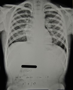
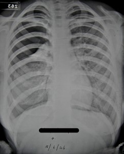
Patient 3/1947 was a 12 year old boy from Lemington-on-Tyne who was admitted to Stannington in January 1947 diagnosed with miliary tuberculosis.  A report on x-ray films taken in December 1946 prior to his admission describes extensive mottled shadowing across both lungs with hilar shadows much enlarged.  The Northumberland County medical officer of health that refers the boy to Stannington gives the following report:

‘States no cough.  Mother says he thinks he gets a bit short of breath at times, and that he has definitely lost weight.  On examination, slight cyanotic tinge; afebrile, pulse 108.  General condition satisfactory (amazing in view of films).  Little made out in chest apart from slight impairment of the air entry at both bases.  Mass of glands at right side of neck.’


Figure 1 is a chest x-ray taken the day after his admission and the report on it simply reads, ‘extensive bilateral miliary spread’.  The extensive mottled ‘snowstorm’ effect is indicative of miliary TB.  Strict bed rest is ordered and at this point he also has an enlarged gland at the angle of the jaw on the right side for which UV light treatment is prescribed.  Over the coming months the abscess on the jaw is described as discharging freely with brownish pus aspirated from it in June 1947.
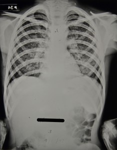
Figure 1 - HOSP/STAN/7/1/2/1371_84
Figure 1 – HOSP/STAN/7/1/2/1371_84
09 Jan 1947
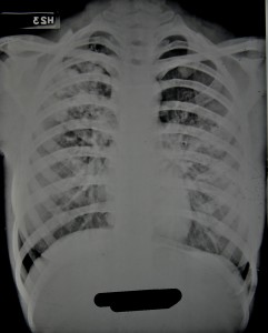
Figure 2 - HOSP/STAN/7/1/2/1371_05
Figure 2 – HOSP/STAN/7/1/2/1371_05
17 Sept 1947


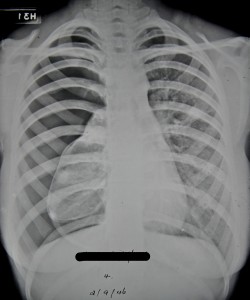
In September 1947 the patient’s doctor suggests that he would be a suitable candidate for streptomycin treatment, which had only recently been introduced at this point.  However, having discussed the case further it was decided that he was not suitable as at this point in time streptomycin was being used for very early cases only and patient 3/1947 by now had a long history of TB and was doing very well without it.  Figure 2, is an x-ray taken around the time streptomycin treatment was being discussed and the report reads, ‘X-ray shows a little improvement.  Each individual lesion is smaller.’


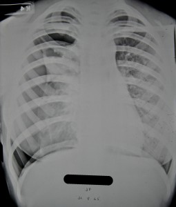
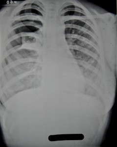
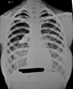
Two months later in November 1947 his condition deteriorates a little and he begins to lose weight and so is again put forward for streptomycin ‘if any available.’  Whilst the attending doctor continues to push for streptomycin over the coming months it is not until November 1948 that the patient receives any.  There are continuing disputes as to whether he is a suitable candidate.  During this time his general condition fluctuates with periods of weight gain and weight loss and x-rays from April and June 1948 show some improvements, figures 3 and 4 respectively.
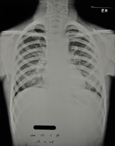
Figure 3 - HOSP/STAN/7/1/2/1371_09
Figure 3 – HOSP/STAN/7/1/2/1371_09
13 April 1948
‘Amazing improvement since last x-ray 3 months ago. The military lesions now appear to be resolving: the apices are almost clear. There is now a more homogenous opacity in the left lower lobe.’
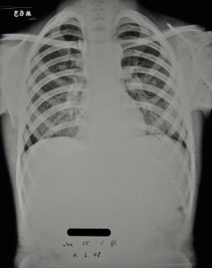
Figure 4 - HOSP/STAN/7/1/2/1371_10
Figure 4 – HOSP/STAN/7/1/2/1371_10
04 June 1948
‘still some mottling at the bases, the apices are clear. The more homogenous density at the L base is getting smaller. Azygos lobe on right side.’


Ultimately the catalyst leading to the decision being made for streptomycin treatment to be commenced appears to be the fact that in June 1948 a swelling over the lower dorsal spine is identified and it is apparent that the tuberculous infection has spread further.  It is clear from radiographic evidence in 1948 that there are three spinal lesions: one in the 5th and 6th dorsal vertebrae causing some kyphosis; another affecting the 1st and 3rd lumbar vertebrae causing some deformity; and a final one in the inferior and anterior part of the body of the 5th lumbar vertebrae with some destruction.


All streptomycin treatment is discontinued by May 1949 when significant improvements in his chest are seen and treatment of the spinal lesions is continued with braces and plaster casts.  He is eventually discharged in July 1950 wearing a spinal brace and continues to be seen by the out-patients’ service until April 1954 when an abscess in the left iliac fossa leads to him being considered for admission to the Queen Elizabeth Hospital.
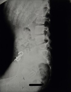
Figure 5 - HOSP/STAN/7/1/2/1371_18
Figure 5 – HOSP/STAN/7/1/2/1371_18
Spinal X-ray, 17 Jan 1950, calcifications in the lungs also evident.
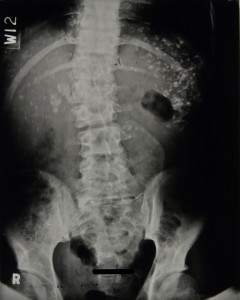
Figure 6 - HOSP/STAN/7/1/2/1371_85
Figure 6 – HOSP/STAN/7/1/2/1371_85
Spinal X-ray, 10 July 1950, 4 days before discharge
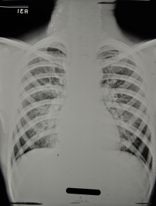
Figure 7 - HOSP/STAN/7/1/2/1371_14 Chest X-ray, 17 Feb 1950, also showing calcifications in neck glands.
Figure 7 – HOSP/STAN/7/1/2/1371_14
Chest X-ray, 17 Feb 1950, also showing calcifications in neck glands.


Sources:

MILLER, F. J. W, SEAL, R. M. E, and TAYLOR, M. D. (1963) Tuberculosis in Children, J & A Churchill Ltd.

Patient 133/1959 – A Case of Fibrocystic Disease

The radiographs held within the Stannington Sanatorium Collection date between 1936 and 1953 and are specific to the period when the sanatorium was used as a hospital for tuberculous children. However, through the process of listing the patient files for Stannington, which continue up until 1966, well into its use as a general children’s hospital, we have uncovered a set of radiographs for one other patient, a 6 year old boy who was admitted to Stannington on 15th June 1959: Patient 133/1959.

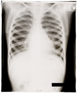
Patient 133/1959 was diagnosed with Fibrocystic Disease of the Pancreas, a generalised hereditary condition amongst children which, despite its name, can affect not only the pancreas but also the liver, lungs and sweat glands and was considered to be the most common cause of chronic non-tuberculous lung disease in children during the 1950s.

This patient was admitted to Stannington with widespread cystic change in both lungs, retarded growth and signs of chronically infected bronchiectasis. His medical notes also refer to ‘finger clubbing’, which involves changes to the areas of soft tissue under and surrounding the finger nails but may also involve the nails themselves. At this stage in his treatment the patient was prescribed penicillin, monitored for changes in weight and subjected to postural drainage.

By September 1959, the boy’s medical notes read:

‘No gain in weight and very little sputum but he has many loud râles in both sides of chest and a loose cough.’


At this time it is also noted that he has been prescribed pancreatin, a drug to help treat the symptoms of cystic fibrosis and to aid with digestion of fat, starch and protein. His lack of weight gain was considered to be associated with problems in the colon and as such his stools were also monitored regularly.

Little change is seen in the notes for the following year, weight gain is still elusive and the patient is often referred to as being small, with râles in the chest and having shortness of breath. However, in October 1960 the severity of this patient’s condition becomes clear as the attending physician writes:

‘Very poor appetite and very difficult.

I do not understand how this problem can be solved.’

However, from May 1961, this boy began to show slow improvements especially within regards to the colon and weight gain is evident with a total gain of 8lbs since admittance. Râles are still noted in the left side of the chest, see lung x-ray in Figure 1.

HOSP-STAN-07-01-02-2243-02
Figure 1 – HOSP-STAN-07-01-02-2243-02

In addition to his medical problems this boy also had learning difficulties according to notes from the hospital school teacher stating he was:

‘very much retarded and would need more individual help and attention ….

He is slow to complete written assignments but makes a good effort. His reading and arithmetic are well below average for his age but he has made satisfactory progress in both within his limited ability.’

As a result the doctors approached the subject of finding this boy a long term residential school for handicapped children to be transferred to following his discharge.

Patient 133/1959, remained in Stannington until 1962, where he continued to make improvements regarding his weight and height and the râles in his chest diminished significantly. In March 1962, he contracted German measles and then later chicken pox in June 1962. Despite these short term illnesses, this patient was discharged home in August 1962, having secured a place at the Windlestone Hall residential school near Ferryhill, County Durham, to begin in September 1962.


Sources:

di Sant’agnese, Paul. A (1955). The Pulmonary Manifestations of Fibrocystic Disease of the Pancreas. Chest. 27(6):654-667

Medline Plus (2015). Clubbing of the Fingers and Toes. http://www.nlm.nih.gov/medlineplus/ency/article/003282.htm

Patient (2015). Pancreatin. http://patient.info/medicine/pancreatin

Surgical Procedures – Artificial Pneumothorax

Pulmonary tuberculosis is by far the most common manifestation of TB witnessed throughout the Stannington records.  Prior to the development and use of any effective antibiotic treatments the most common form of intervention was the induction of an artificial pneumothorax.  Many of the different treatments employed to treat TB of all types at this time were based on the principles of resting and isolating the affected area, and the thinking behind artificial pneumothorax treatment was no different.


A needle would be inserted through the chest wall to allow for the insertion of air into the pleural cavity.  The amount of air inserted would depend on the size of the patient as well as how much the physician in charge though the patient could realistically manage in one go and how quickly they wished the lung to collapse.  Once inserted the pressure from the air would force the lung to collapse in on itself and to cease functioning properly.  The entire lung would not necessarily be collapsed at once, either because it wasn’t necessary for treatment or because fibrotic adhesions between the lung and the chest wall as a result of the disease prevented it from doing so.  Where only part of the lung was affected it would not be desirable to collapse the whole lung and in such instances just one lobe might be collapse.  Bilateral artificial pneumothorax was also a possibility, whereby part of both lungs would be collapsed at the same time.  A state of collapse could be maintained for a period of months or even years and required the patient to undergo regular refills of air in order to do so.


A great number of radiographic illustrations of the progression of a collapse are available in the Stannington collection.  One patient, 2/1946, has a large amount of radiographs taken over a period of two years which demonstrate the change in the lung from admission and through the progressive stages of lung collapse.


Patient 2/1946 was female an age15 when she was admitted to Stannington on 21 June 1945 with pulmonary TB stage 3, at which point her sputum tested positive for TB also.  A report on an x-ray taken pre-admission reads:

‘Right lung shows several active foci beginning to coalesce.  There is extensive infiltration in the upper zone & suspicious blotchy areas in the middle zone.  A small calcified opacity in the right lower zone.  The left lung shows infiltration in the middle zone.  The upper zone and apex are clear.  Early active foci are noticeable in both lungs in the affected areas.’

Figure 1 was the first x-ray taken after admission on 25 June 1945 being three weeks later than the one reported above.  Observations on this x-ray note:

‘Scattered foci in right upper zone.  One definite cavity.  Increased bronchial marking at both bases.’

HOSP/STAN/7/1/2/1057_22 25 June 1945
Figure 1 – HOSP/STAN/7/1/2/1057_22
25 June 1945
HOSP/STAN/7/1/2/1057_18 31 Aug 1945
Figure 2 – HOSP/STAN/7/1/2/1057_18
31 Aug 1945


It was quickly decided that and artificial pneumothorax should be induced on the right side and this took place on 16 Aug 1945. Figure 2 taken later on that month shows the initial results of the artificial pneumothorax.  The black area along the lateral side of the right lung is evidence of the air that has been inserted and the lung has begun to compress.


The collapse was maintained well into 1947 which involved her having refills of air every two weeks throughout this period.  For the first three months she received refills of 200-300ccs of air at a time, progressing to 400ccs the month after, and then eventually 500-600ccs at a time.  Figures 3-6 show the progression of the artificial pneumothorax as more air is inserted and the lung collapses further.  Over time we can see that the cavity in the right mid zone collapses and closes, one of the main aims of the treatment.  In early June 1946 a procedure was performed to divide adhesions between the lung and the chest wall which allowed the collapse to progress further.  She was discharged in June 1947 with her condition described as improved.


Figure 3 - HOSP/STAN/7/1/2/1057_23 17 Jan 1946
Figure 3 – HOSP/STAN/7/1/2/1057_23
17 Jan 1946
Figure 4 - HOSP/STAN/7/1/2/1057_09 18 June 1946
Figure 4 – HOSP/STAN/7/1/2/1057_09
18 June 1946


Figure 5 - HOSP/STAN/7/1/2/1057_10 2 Sept 1946
Figure 5 – HOSP/STAN/7/1/2/1057_10
2 Sept 1946
Figure 6 - HOSP/STAN/7/1/2/1057_27 15 April 1947
Figure 6 – HOSP/STAN/7/1/2/1057_27
15 April 1947