Patient Files

As part of the Stannington collection we have patient case files spanning the years 1939-1966 containing a wealth of medical and social information to support that found in the radiographs.  The earlier files have a different format to the later ones owing to a change in the administration of patient records at Stannington which occurred in 1946.  Up to 1946 the patient records take a much larger format and the patients were all allocated their own unique patient number based on their date of discharge, whereas from 1946 onwards standard sized paper files come into use with patient numbers being based on date of admission.

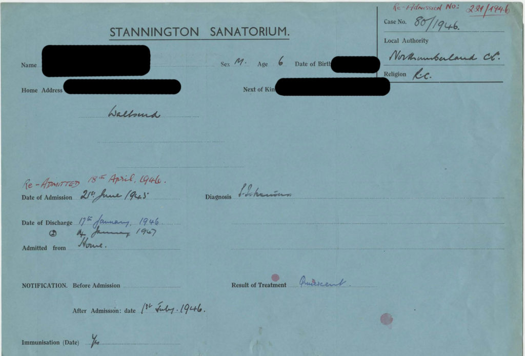
[HOSP/STAN/7/1/1/1587]

The above file is an example of one of the later files with the patient’s name redacted for confidentiality purposes.  Three different colour files were used, each one indicating the type of tuberculosis the patient was suffering from.  Blue files were used for sufferers of pulmonary TB, pink files for non-pulmonary TB, and finally green files for TB of the bones and joints.  This image gives a good indication of the sort of information that can be found on the files, which is also indicative of the information we will be recording in our catalogue.  The information featured in the catalogue for each patient will be as follows: patient number, date of admission, date of discharge, sex, age on admission, home town, diagnosis, result of treatment, where admitted from, the local authority sending them, and where applicable any re-admission numbers and dates.


To clarify some of the information given on the file, the date of immunisation refers to immunisation against diphtheria, not tuberculosis, as widespread vaccination against TB was not yet in place.  As a contagious disease and a major concern for public health, all diagnosed cases of tuberculosis had to be made known to the local public health authorities, which is what the notification date refers to.


Each case of tuberculosis had to be classified according to centrally issued guidelines and this is often noted on the patient’s file under diagnosis. The first distinction made when classifying the disease is between pulmonary and non-pulmonary tuberculosis, with pulmonary including TB of the pleura and intrathoracic glands and any patient suffering from a combination of pulmonary and non-pulmonary TB would be classified as pulmonary.  Cases of pulmonary TB could then be subdivided between TB minus and TB plus.  Cases in which tubercle bacilli have never been found in the sputum or other pathology samples are classed as TB minus, as the above patient is.  TB plus on the other hand applies to cases in which tubercle bacilli have at some point been found and is subdivided further into 3 groups; group 1 applying to cases with slight constitutional disturbances if any and limited physical signs, group 3 for cases showing profound constitutional disturbance or deterioration and with little or no prospect of recovery, and finally group 2 for all cases which cannot be placed within groups 1 or 3.  Patients suffering from non-pulmonary tuberculosis would be classified according to the site of the lesion, for example, tuberculosis of the bones and joints, abdominal tuberculosis, tuberculosis of other organs, and tuberculosis of the peripheral glands.


There is also a space on each patient file to enter the result of treatment and there were also central guidelines covering this.  Most patients leaving Stannington are deemed to be ‘quiescent’, meaning that they have no signs or symptoms of tuberculous disease and any sputum is free of tubercle bacilli.  A patient’s condition could also be classed as ‘arrested’ by which it is meant that in pulmonary cases the disease has been quiescent for at least two years and in non-pulmonary cases it is quiescent and there is no reason to believe it will recur.  And finally, a patient could be considered to be ‘recovered’ if the disease had been arrested for at least three years.


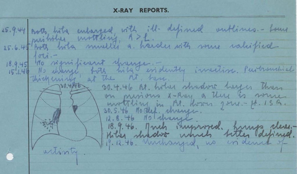
The information that you can expect to find within the patient records does vary from patient to patient but generally includes data on other family members, living conditions, medical history, temperature charts, x-ray reports, pathology reports, details of progress, and any correspondence with family members or local authorities.  The correspondence contained within the files can give a fascinating insight into social problems and the impact tuberculosis could have on families at the time adding an extra dimension to the medical information that we expect to find.  The image below is an example of the x-ray reports that can be found in the back of some of the files; it is quite common for files dating from the mid-1940s to find small diagrams of what was seen in the x-rays also.


x-ray reports

[HOSP/STAN/7/1/1/1587]


Case Study – Pulmonary Tuberculosis

In this post we’re going to explore the progression of pulmonary tuberculosis in one particular patient from Stannington Sanatorium in order to gain an insight into some of the common approaches to the treatment of the disease at this time.


Patient 95/1947 was admitted to Stannington Sanatorium on 4th September 1947 at the age of 12. After having begun to feel ill earlier in the year she was examined at the local clinic and sent for x-ray whereupon it was determined that she should be admitted to the sanatorium for treatment.  Prior to admission she had been living with her mother, step-father, two younger brothers and one younger sister in a 3 roomed house in Cockermouth which had no inside water or inside toilet.  The only family history of TB had been her father who had died from the disease when she was still a baby.  On admission she had no cough but a very poor appetite and was losing weight, weighing only 4st 0lbs 6oz.  There were no other physical symptoms or abnormalities reported.


The report on her first x-ray taken 4 days after admission reads:

Tuberculous infiltration of both upper lobes with a large cavity in the mid-zone & a smaller one at the left apex.  There are several small calcified foci in the right upper lobe.”

Continuing reports over the next 4 months describe great improvement on the right side with the cavity in the right mid-zone no longer being visible.  However, the condition of the left side continues to deteriorate with a report 7 months after admission stating that the “cavity in the left upper lobe is now very much larger 1 ½” in diameter.”


NRO-3000-HOSP-STAN-07-01-02-1444-19

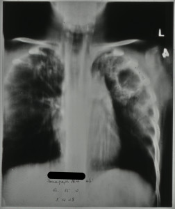
[HOSP/STAN/7/1/2/1444/19 – tomograph showing large cavity in left upper lobe, Dec 1948]


During her stay a series of different treatments were attempted to reduce the cavities.  Two months after admission in November 1947 her doctor initially observed that it was “doubtful if a satisfactory collapse could be obtained.  No treatment recommended.  Outlook very poor.”  Nevertheless, two months later in January an artificial pneumothorax was attempted but without success.


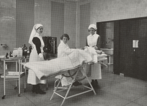
Artificial pneumothoraxes were performed on patients with the intention of resting the affected lung and hopefully collapsing the cavities at the same time whilst preventing any further spread as a collapsed lung was less likely to spread bacilli.  The procedure had been shown to effect a marked improvement in the size of tuberculous cavities for some patients but could at the same time be a dangerous procedure with a risk of air embolisms, pleural shock, sepsis, emphysema and effusion.


HOSP_STAN_9_1_1

 [HOSP/STAN/9/1/1, artificial pneumothorax treatment being performed in Stannington]


Three months later, after observing the growth of the cavity in the left upper lobe, a phrenic crush followed by a pneumoperitoneum was recommended and she was transferred to Shotley Bridge Hospital soon after for the procedures to be performed.  By crushing the left phrenic nerve, situated in the neck, they would be able to disable the left diaphragm thus forcing the muscle to relax and lift up, with the idea being that this would then rest the lower part of the lung.   A pneumoperitoneum was often performed in conjunction with the phrenic crush and involved inserting air into the abdominal peritoneal cavity forcing the diaphragm up.


Unfortunately after the patient was transferred to Shotley Bridge Hospital for the above procedures she never returned to Stannington and so we do not have any later case notes to follow up the result of her treatment.  However, some later correspondence does tell us that she was moved to Poole Sanatorium from where she was eventually discharged in May 1950.


The surgical procedures described here sound very drastic from a modern perspective but were a common approach in the pre-antibiotic era.  With no effective drug treatments surgical approaches such as these were at the forefront of tuberculosis treatment and looking through the files of Stannington Sanatorium it is clear that many of their young patients recovered, or at least showed significant improvements, and went on to live normal lives.

Types of Tuberculosis at Stannington Sanatorium

Tuberculosis is a bacterium based infectious disease, known as Mycobacterium-tuberculosis. It is a widely held misconception that tuberculosis (TB) is a disease that only affects the lungs. In fact TB can affect any part of the body, both the skeleton and the soft tissue. The key aim here is to identify the various types of TB that affect children and how these affect different parts of the body, with a view to making the patient records and radiographs of the Stannington Sanatorium more understandable.

Primary Infection

Tuberculosis is generally contracted through the inhalation of infected droplets, usually the result of a cough or sneeze, from an individual with active infection. Once inhaled the infection enters the lungs where it manifests itself as a primary complex (Primary TB). Initial infection can be identified by a fever and night sweats which would last up to two weeks, not dissimilar to the effects of pneumonia. This may be followed by skin dullness, weight loss, an increased respiratory rate and haemoptysis (coughing up blood or blood stained sputum). The primary infection goes into a state of incubation during which it multiplies on a small scale to reduce the immune system’s ability to identify its presence, it then spreads throughout the body.

Secondary Infection

There are two ways in which tuberculosis can be disseminated throughout the body, through the lymphatic system and through haematogenous spread (blood stream). The process of widespread dissemination of infection is categorised as miliary tuberculosis, which occurs early in primary infection. It is identified as a series of tiny spots in radiographs, but also causes Chorodial tubercles or pale lesions on the optic nerve which serve as an important symptom in diagnosing this form of TB, particularly in children. Due to its disseminated nature within the body, miliary TB is responsible for a number of other forms of secondary tuberculosis. Organs including the liver, spleen, kidneys, bladder and genitalia are all affected by tuberculosis which are connected to haematogenous miliary TB.

Miliary tuberculosis is also closely linked with the most fatal form of TB, tuberculosis-meningitis. Affecting the central nervous, TB-meningitis has been attributed with a large proportion of tuberculosis related deaths amongst children. It is caused when tuberculosis bacteria infiltrate the fluids surrounding the brain and spinal cord causing small abscesses, which when burst cause tuberculosis-meningitis. It is often identified from symptoms such as irritability, listlessness, headaches and vomiting.

Tuberculosis also manifests itself within the skeletal system. Tuberculosis of the bones and joints is more prevalent in children, and is well represented in the patients of the Stannington Sanatorium. Spinal involvement in tuberculosis, also known as Pott ’s disease, is considered to account for the highest number of skeletal tuberculosis cases. Tuberculosis affects the spine through the destruction of the discs between individual vertebrae and ultimately the collapse of the spinal column causing an identifiable curvature of the spine. The compression or angular result of spinal tuberculosis can proceed to cause paraplegia and/or neurological damage depending on the number and location of the vertebrae involved.

Extra-spinal tuberculosis can be divided into two categories, although they commonly fall under the title ‘bone and joint TB’, the first being tuberculosis-osteomyelitis. The effect of this form of TB is usually characterised by destruction of portions of the long bones and the epiphyses (ends of the long bones), followed by periostitis, the process of new bone growth. In the hands and feet, there is also usually noticeable swelling of the soft tissue in addition to bony anomalies.

The second form of skeletal tuberculosis is tuberculosis-arthritis, which affects the joints, including the hip, knee and elbow. This can be caused by direct invasion from adjoining bones with tuberculosis-osteomyelitis or through miliary TB. Tuberculosis of the joint causes erosion of the joint surface and depletion in the space between the joint. This can subsequently cause septic arthritis of the joint, in which the joint surface is destroyed ultimately causing ankylosis (stiffness and immobility).

Abdominal-tuberculosis is another common extra-pulmonary form of tuberculosis. This was considered a primary form of tuberculosis prior to milk pasteurisation, as individuals could contract the bovine strain of mycobacterium-tuberculosis (mycobacterium-bovis) through the ingestion of contaminated meat or unpasteurised milk. It is characterised by ulcers and thickening of the bowel wall and can lead to the secondary tuberculosis: peritoneal-tuberculosis. Abdominal tuberculosis often healed spontaneously sometimes with calcifications in the abdominal region which act as radiographic evidence of the disease.

Tuberculosis of the pleural cavity, the space found between two layers of pleura surrounding the lung itself, is considered to be a direct complication of primary infection of the lungs. Pleurisy with effusion and empyema are the two most common forms of tuberculosis found within the pleural cavity. Despite the involvement of primary infection within the lungs and the pleural cavity, tuberculosis of the pleural space is considered as an extra-pulmonary form of tuberculosis.

A less common form of TB is tuberculosis of the skin, identifiable by the nodular skin lesions found on the face; the most common form of this is lupus vulgaris.

Finally chronic pulmonary tuberculosis, also known as tertiary, re-infected or adult tuberculosis, where infected bacteria encounter an area already sensitised to the infection. This can occur in children who have had already had primary tuberculosis and usually appears in adolescence and early adulthood.

Stannington Sanatorium

The diagnoses of patients from Stannington Sanatorium display a range of different types of tuberculosis. Pulmonary case files are the most numerous, however these also consist of those patients admitted for non-TB pulmonary conditions such as bronchiectasis. Still once all non-TB patients are removed from the case files, pulmonary or primary TB accounts for a significant proportion of patients treated at the Stannington Sanatorium.

All patients were subject to a chest radiograph upon admission as a diagnostic tool. As a result the radiographic collection holds a predominance of chest radiographs displaying a range of chest related forms of tuberculosis and non-tuberculosis conditions.

Miliary tuberculosis features prominently within the patient files, and is usually associated with those suffering from a severe form of the disease. As an extension of this form of tuberculosis there are also a range of organ, nerve, skin and skeletal related tuberculosis cases.

Stannington also offers a generous proportion of bone and joint tuberculosis allowing for detailed examination of the ways different bones were affected by the infection. This allows for the study of skeletal progression of the disease, particularly useful for comparative purposes when looking at tuberculosis in archaeological collections.


Sources

Meningitis Research Foundation: http://www.meningitis.org/disease-info/types-causes/tb-meningitis

Harisinghani, M.G; McLoud, T.C; Shepard, J.O; Ko, J.P; Shroff, M.M; Mueller, P.R (2000). Tuberculosis from Head to Toe, in Radiographics pp.449-70. http://www.ncbi.nlm.nih.gov/pubmed/10715343