World Tuberculosis Day 2016!

In honour of World Tuberculosis Day, we have a guest blog from Rebecca Cessford. Rebecca is a PhD researcher with the AHRC funded Heritage Consortium based at the Universities of Hull and Bradford. She will be using the Stannington Sanatorium Collection to study tuberculosis in the past using the archaeology of human remains and medical history. Here she tells us about her research and the role of the Stannington Collection in it.


When we think of tuberculosis (TB), images are conjured of a romantic disease causing a bloody cough, a pale complexion and weight-loss, the romanticised disease of the 19th century. What we do not think of is TB roaming the streets today. But tuberculosis is still a great threat, with over a million people dying of the disease each year and over 6,500 new cases declared in the UK during 2014. With increasing multidrug resistant strains of tuberculosis, is it possible to look back at a time before antibiotic drug therapy to better understand the future of this global emergency?

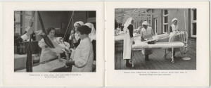
Early Discovery, Early Recovery 1929. Image from the National Library of Medicine, USA


Tuberculosis is a disease that extends as far back as the Neolithic period in Europe, with the earliest case reported in England coming from Dorset dating to the Iron Age. However, our ability to identify tuberculosis in skeletal remains from archaeological contexts is difficult. Firstly, tuberculosis of the bones and joints only affects 3-5% of all cases. Secondly, bone can only react to disease in a limited number of ways with many diseases causing similar bony destruction and remodelling. There are also problems identifying tuberculosis in the remains of children, due mainly to the under-representation of children in the archaeological record.

The most characteristic feature of tuberculosis in the skeleton is Pott’s Spine, an angular deformity in the mid to lower spinal column caused by the collapse of one or more vertebral bodies. The presence of this deformity has, for many years, been the only way of diagnosing tuberculosis in human remains with any certainty, despite the fact that any bone in the body can be affected. Advances in ancient DNA and biomolecular studies in archaeology mean tuberculosis can be tested for, even in the absence of any physical pathologies. However, these destructive and costly procedures are not without their limitations, still leaving much reliance on routine macroscopic observations (seen with the naked eye) of dry bone remains.
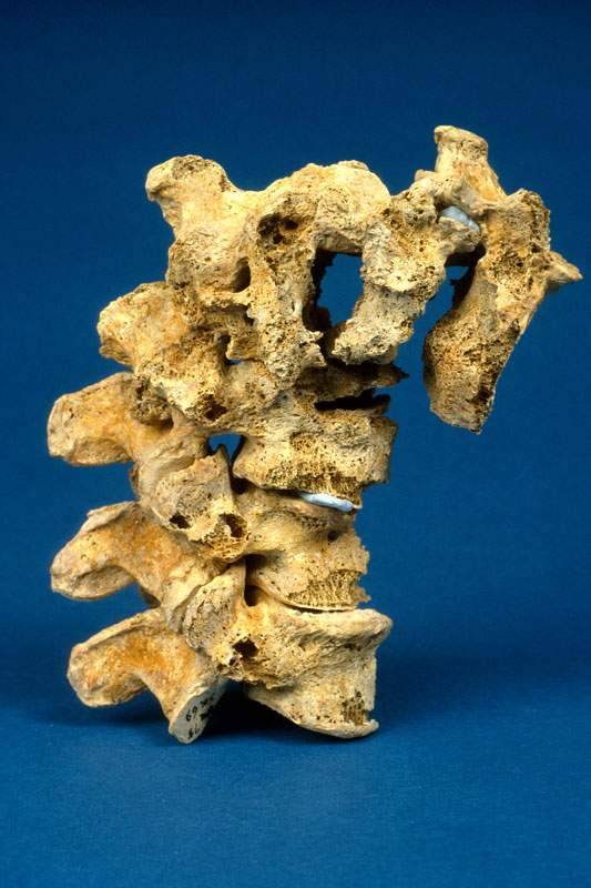
Pott's Spine the main diagnostic feature of tuberculosis in skeletal remains. Image courtesy of https://www.dur.ac.uk/images/archaeology/researchprojects/Roberts_TB.jpg
Pott’s Spine – characteristic collapse of the vertebral bodies causing an angular deformity of the spine due to tuberculosis. Image from University of Durham.


My research aims to look at the potential for using pre-antibiotic clinical radiographs (x- rays) as an aid to the macroscopic identification of tuberculosis in human remains, focussing specifically on infants and children. To do this, I intend to undertake a thorough examination of all the radiographs demonstrating skeletal tuberculosis to look at variations in progression of disease over time; the outcomes of healing on bones and the distribution of tuberculosis across the body where more than one bone was involved. In addition to this I will look at the corresponding medical file for each set of radiographs drawing on details outlined in the medical notes and x-ray reports to add to my own observations from the radiographs for an informed review of the underlying processes to bone and soft tissue being observed. It is hoped that the compilation of this data will provide a more detailed understanding of the processes involved in advancing tuberculous infection with comparative examples from pre-antibiotic radiographs. This strives to increase the ability to diagnose tuberculosis in archaeological remains even in the absence of Pott’s Spine.
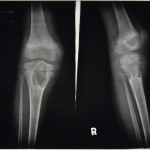
 

Tuberculosis of the Knee: HOSP-STAN-07-01-02-91_09
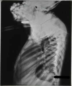
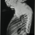
Tuberculosis of the Spine - HOSP-STAN-07-01-02-1662-22
Tuberculosis of the Spine – HOSP-STAN-07-01-02-1662-22
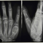
Tuberculosis affecting the finger bones: HOSP-STAN-07-01-02-641_07


By studying the patterns of tuberculosis in the past we are better informed when it comes to dealing with the disease in the present and in the future. To be able to offer an evidence-based and informed approach to tackling tuberculosis we need better criteria for diagnosing it macroscopically in archaeological human remains, to get a more encompassing view of the various manifestations associated with it. The outcomes of my research will aim to act as an aid to the identification and study of tuberculosis in children in relation to archaeological remains further identifying the worth of pre-antibiotic medical records.

The Stannington Collection is a unique resource for studying this long standing infectious disease in children from the early to mid-20th century, many of which are still alive today living with the memories and/or side effects of the disease. I would also like to take this opportunity to thank the former patients of Stannington Sanatorium who expressed support for academic research to be undertaken on the collection during the first phase of the Stannington Sanatorium Project; their support makes research all the more worthwhile.

Pott’s Disease (Tuberculosis of the Spine)

The spine is the most frequent site of skeletal involvement in tuberculosis of the bones and joints. Commonly known as Pott’s Disease, after Sir Percival Pott who first described the condition in 1779, tuberculous osteomyelitis of the spine affects between 25 and 60% of all individuals suffering from skeletal tuberculosis. It is most commonly seen in children and young adults, predominantly affecting the thoracic and upper lumbar regions of the spine, although evidence of cervical involvement also exists. Spinal lesions begin in the cartilage between the vertebrae or in one or more vertebral bodies, this leads to a narrowing of the joint space, noticeable in radiological examination. Paravertebral abscesses can also occur when diseased tissue in the vicinity of the affected vertebrae forms a mass and pus collects. With the expansion of this abscess there can be a loss of blood supply to the vertebral body resulting in a loss of integrity causing the vertebral column to collapse creating an angulation or ‘kyphosis’ to the spine. The collapsed vertebrae form a wedge, known as a ‘Gibbus deformity’, which can lead to compression of the spinal cord resulting in paraplegia, as well as functional problems with the pulmonary system.

There are numerous cases of spinal tuberculosis in the records from Stannington, all varying in their severity and final outcome. Below are two examples of the different types of spinal tuberculosis and the methods used to treat it.


Case Study 1 – Dorsal (Thoracic) Spine

Patient 17/1949, a 4 year old boy, was transferred to Stannington Sanatorium from the Royal Victoria Infirmary (RVI) in January 1949. His medical history had included a bout of pertussis, whooping cough, complicated by pneumonia followed a year later by lethargy and a swollen knee. In April 1947 he was admitted to Earl’s House Sanatorium with a primary tuberculous complex in the left upper zone of his chest and TB of the left upper tibial epiphysis and upper dorsal (thoracic) spine.


Paraplegia developed in September 1948 and he was transferred to the RVI that December showing signs of wasting and obvious kyphosis in the upper dorsal spine with paraplegia evident and total incontinence. His notes state that his head and thorax were encased in plaster cast, as was the left leg, to immobilise the affected areas. The incontinence was dealt with by applying a tube. Tuberculosis of the spine was relatively advanced, with the 3rd and 4th dorsal vertebrae having collapsed resulting in a noticeable kyphosis, seen in Figure 1.

FIGURE 1: HOSP-STAN-07-01-02-1662-22

On admission to Stannington it is recorded that the radiographs showed a high dorsal lesion. The plaster cast encasing the head and thorax was removed and the patient was fixed to a short plaster boat with head piece, see left image in Figure 4.

FIGURE 4: HOSP-STAN-07-01-02-1662-24
FIGURE 3: HOSP-STAN-07-01-02-1662-24
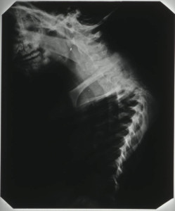
FIGURE 2: HOSP-STAN-07-01-02-1662-16


Radiographic images show further kyphosis, Figure 2, and the collapse of the vertebral bodies. Porosity is evident in the vertebral bodies in the upper dorsal region, identifiable by their translucent nature in Figure 3, giving rise to the extent of the infection.

In November 1949, the patient was fitted for a plastic splint. This was to fit

‘from the hips up the trunk extending over the neck to the occiput, reinforced with metal where necessary’.

The spinal lesion was considered quiescent by April 1952, all evidence of paraplegia having cleared up. However, he was to be fitted with a splint with a shaped head piece to immobilise the spine as much as possible. This patient was discharged in March 1953 and his brace discarded entirely in May 1953. He continued to be seen as an outpatient until February 1959.

HOSP-STAN-09-01-01
Figure 4:  HOSP-STAN-09-01-01


Case Study 2 – Cervical Vertebrae

Patient 148/1948, a 3 year old girl, was initially admitted in January 1948 (Patient Number 8/1948) with a Primary Tuberculous Complex of the right mid-zone.

Preliminary medical reports described this girl as having had an enlarged right hilar shadow, a shadow of the hilar lymph nodes, and ‘shotty,’ swollen, glands with an impetigious lesion on the scalp. However, her initial stay at Stannington was short as she was removed against medical advice by her mother 28 days after admittance, only to be re-admitted seven months later with TB of the cervical spine.

Following an examination by the surgeon, Mr Stanger, on re-admission a comprehensive outline of her condition was given:

The lower surface of the 2nd c.v (cervical vertebrae) is involved; the body of the 3rd c.v is completely destroyed and the upper surface of the 4th is probably eroded.

This child should have every bone in her body x-rayed.

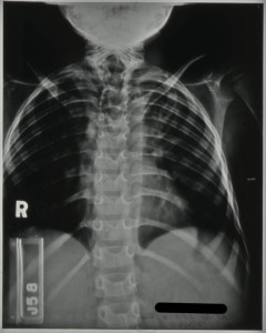
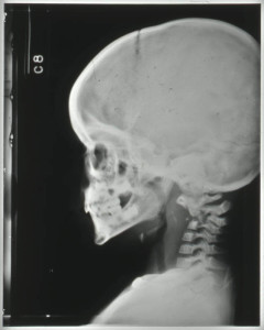
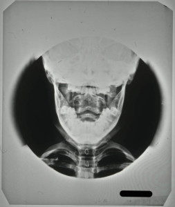
The destruction of the vertebral bodies can be seen in the radiographs in Figures 5 and 6. Figure 5 shows the collapse of the vertebrae inwards creating a wedged shape in the neck. Figure 6, taken through the open mouth of the patient in order to gain a clear veiw of the vertebral bodies of the cervical vertebrae in the neck, shows a loss in denisty and clearly defined outer edges of the vertebral bodies due to collapse.

FIGURE 4: HOSP-STAN-07-01-02-1625-14
FIGURE 5: HOSP-STAN-07-01-02-1625-14
FIGURE 6: HOSP-STAN-07-01-02-1625-43


It is likely the request for all bones in her body to be x-rayed came from the suspicion that other areas of the skeleton had been affected by the disease. The request was carried out with the x-ray report card indicating that anteroposterior (AP) and lateral radiographs, where possible, were taken of the chest, spine, legs and hips. The patient was immobilised on a Bradford frame, a rectangular metal frame with canvas straps to hold the individual in a prone or supine position, seen in the right hand image of Figure 4.

Between September and December 1948 the patient is noted to have developed a number of additional symptoms, including vomiting sputum; patchy erythema (a scarlet rash) on her chest; purulent nasopharyngeal discharge (discharging pus from the nose); aural discharge; an inflamed throat and enlarged cervical glands.

By July 1949, these symptoms had largely been addressed and the patient was showing improvement. Immobilisation was considered satisfactory as a form of treatment and a moulded plastic splint was to be prepared for the patient, to consist of

a jacket taken from the hips and extending upwards to embrace the head and the occipital region to the chin.

This was later described as being reinforced with steel both vertically and transversely.


One year later, further examination by Mr Stanger noted that the disease had involved the 2nd, 3rd and 4th cervical vertebrae; the bodies of the 2nd and 3rd were showing signs of fusion and bone regeneration. It is at this point in July 1950, two years after first being admitted, that the child was allowed to ‘get up’.


This patient was discharged in December 1950, as being clinically and radiologically inactive and able to dispense with the splint. She continued to be seen as an outpatient at Stannington until 1956. Her last out-patient report stating that there was no deformity and no limitation of movement. Sound fusion was noted between both the vertebral bodies and posterior articulation of the 2nd, 3rd and 4th cervical vertebrae.


Further radiographic images can be seen on the Stannington Sanatorium ‘Radiographs from Stannington’ Flickr stream https://www.flickr.com/photos/99322319@N07/sets/72157648833066476/


Sources

Miller, F.J.W, Seal, R.M.E and Taylor, M.D (1963). Tuberculosis in Children. J & A Churchill Ltd.

Roberts, C and Buikstra, J (2003). The Bioarchaeology of Tuberculosis: A Global View on Reemerging Disease. Univesity Press of Florida.

Roberts, C and Manchester, K (2006). Archaeology of Disease (3rd Edition). Cornell University Press.

Types of Tuberculosis at Stannington Sanatorium

Tuberculosis is a bacterium based infectious disease, known as Mycobacterium-tuberculosis. It is a widely held misconception that tuberculosis (TB) is a disease that only affects the lungs. In fact TB can affect any part of the body, both the skeleton and the soft tissue. The key aim here is to identify the various types of TB that affect children and how these affect different parts of the body, with a view to making the patient records and radiographs of the Stannington Sanatorium more understandable.

Primary Infection

Tuberculosis is generally contracted through the inhalation of infected droplets, usually the result of a cough or sneeze, from an individual with active infection. Once inhaled the infection enters the lungs where it manifests itself as a primary complex (Primary TB). Initial infection can be identified by a fever and night sweats which would last up to two weeks, not dissimilar to the effects of pneumonia. This may be followed by skin dullness, weight loss, an increased respiratory rate and haemoptysis (coughing up blood or blood stained sputum). The primary infection goes into a state of incubation during which it multiplies on a small scale to reduce the immune system’s ability to identify its presence, it then spreads throughout the body.

Secondary Infection

There are two ways in which tuberculosis can be disseminated throughout the body, through the lymphatic system and through haematogenous spread (blood stream). The process of widespread dissemination of infection is categorised as miliary tuberculosis, which occurs early in primary infection. It is identified as a series of tiny spots in radiographs, but also causes Chorodial tubercles or pale lesions on the optic nerve which serve as an important symptom in diagnosing this form of TB, particularly in children. Due to its disseminated nature within the body, miliary TB is responsible for a number of other forms of secondary tuberculosis. Organs including the liver, spleen, kidneys, bladder and genitalia are all affected by tuberculosis which are connected to haematogenous miliary TB.

Miliary tuberculosis is also closely linked with the most fatal form of TB, tuberculosis-meningitis. Affecting the central nervous, TB-meningitis has been attributed with a large proportion of tuberculosis related deaths amongst children. It is caused when tuberculosis bacteria infiltrate the fluids surrounding the brain and spinal cord causing small abscesses, which when burst cause tuberculosis-meningitis. It is often identified from symptoms such as irritability, listlessness, headaches and vomiting.

Tuberculosis also manifests itself within the skeletal system. Tuberculosis of the bones and joints is more prevalent in children, and is well represented in the patients of the Stannington Sanatorium. Spinal involvement in tuberculosis, also known as Pott ’s disease, is considered to account for the highest number of skeletal tuberculosis cases. Tuberculosis affects the spine through the destruction of the discs between individual vertebrae and ultimately the collapse of the spinal column causing an identifiable curvature of the spine. The compression or angular result of spinal tuberculosis can proceed to cause paraplegia and/or neurological damage depending on the number and location of the vertebrae involved.

Extra-spinal tuberculosis can be divided into two categories, although they commonly fall under the title ‘bone and joint TB’, the first being tuberculosis-osteomyelitis. The effect of this form of TB is usually characterised by destruction of portions of the long bones and the epiphyses (ends of the long bones), followed by periostitis, the process of new bone growth. In the hands and feet, there is also usually noticeable swelling of the soft tissue in addition to bony anomalies.

The second form of skeletal tuberculosis is tuberculosis-arthritis, which affects the joints, including the hip, knee and elbow. This can be caused by direct invasion from adjoining bones with tuberculosis-osteomyelitis or through miliary TB. Tuberculosis of the joint causes erosion of the joint surface and depletion in the space between the joint. This can subsequently cause septic arthritis of the joint, in which the joint surface is destroyed ultimately causing ankylosis (stiffness and immobility).

Abdominal-tuberculosis is another common extra-pulmonary form of tuberculosis. This was considered a primary form of tuberculosis prior to milk pasteurisation, as individuals could contract the bovine strain of mycobacterium-tuberculosis (mycobacterium-bovis) through the ingestion of contaminated meat or unpasteurised milk. It is characterised by ulcers and thickening of the bowel wall and can lead to the secondary tuberculosis: peritoneal-tuberculosis. Abdominal tuberculosis often healed spontaneously sometimes with calcifications in the abdominal region which act as radiographic evidence of the disease.

Tuberculosis of the pleural cavity, the space found between two layers of pleura surrounding the lung itself, is considered to be a direct complication of primary infection of the lungs. Pleurisy with effusion and empyema are the two most common forms of tuberculosis found within the pleural cavity. Despite the involvement of primary infection within the lungs and the pleural cavity, tuberculosis of the pleural space is considered as an extra-pulmonary form of tuberculosis.

A less common form of TB is tuberculosis of the skin, identifiable by the nodular skin lesions found on the face; the most common form of this is lupus vulgaris.

Finally chronic pulmonary tuberculosis, also known as tertiary, re-infected or adult tuberculosis, where infected bacteria encounter an area already sensitised to the infection. This can occur in children who have had already had primary tuberculosis and usually appears in adolescence and early adulthood.

Stannington Sanatorium

The diagnoses of patients from Stannington Sanatorium display a range of different types of tuberculosis. Pulmonary case files are the most numerous, however these also consist of those patients admitted for non-TB pulmonary conditions such as bronchiectasis. Still once all non-TB patients are removed from the case files, pulmonary or primary TB accounts for a significant proportion of patients treated at the Stannington Sanatorium.

All patients were subject to a chest radiograph upon admission as a diagnostic tool. As a result the radiographic collection holds a predominance of chest radiographs displaying a range of chest related forms of tuberculosis and non-tuberculosis conditions.

Miliary tuberculosis features prominently within the patient files, and is usually associated with those suffering from a severe form of the disease. As an extension of this form of tuberculosis there are also a range of organ, nerve, skin and skeletal related tuberculosis cases.

Stannington also offers a generous proportion of bone and joint tuberculosis allowing for detailed examination of the ways different bones were affected by the infection. This allows for the study of skeletal progression of the disease, particularly useful for comparative purposes when looking at tuberculosis in archaeological collections.


Sources

Meningitis Research Foundation: http://www.meningitis.org/disease-info/types-causes/tb-meningitis

Harisinghani, M.G; McLoud, T.C; Shepard, J.O; Ko, J.P; Shroff, M.M; Mueller, P.R (2000). Tuberculosis from Head to Toe, in Radiographics pp.449-70. http://www.ncbi.nlm.nih.gov/pubmed/10715343