Pott’s Disease (Tuberculosis of the Spine)

The spine is the most frequent site of skeletal involvement in tuberculosis of the bones and joints. Commonly known as Pott’s Disease, after Sir Percival Pott who first described the condition in 1779, tuberculous osteomyelitis of the spine affects between 25 and 60% of all individuals suffering from skeletal tuberculosis. It is most commonly seen in children and young adults, predominantly affecting the thoracic and upper lumbar regions of the spine, although evidence of cervical involvement also exists. Spinal lesions begin in the cartilage between the vertebrae or in one or more vertebral bodies, this leads to a narrowing of the joint space, noticeable in radiological examination. Paravertebral abscesses can also occur when diseased tissue in the vicinity of the affected vertebrae forms a mass and pus collects. With the expansion of this abscess there can be a loss of blood supply to the vertebral body resulting in a loss of integrity causing the vertebral column to collapse creating an angulation or ‘kyphosis’ to the spine. The collapsed vertebrae form a wedge, known as a ‘Gibbus deformity’, which can lead to compression of the spinal cord resulting in paraplegia, as well as functional problems with the pulmonary system.

There are numerous cases of spinal tuberculosis in the records from Stannington, all varying in their severity and final outcome. Below are two examples of the different types of spinal tuberculosis and the methods used to treat it.


Case Study 1 – Dorsal (Thoracic) Spine

Patient 17/1949, a 4 year old boy, was transferred to Stannington Sanatorium from the Royal Victoria Infirmary (RVI) in January 1949. His medical history had included a bout of pertussis, whooping cough, complicated by pneumonia followed a year later by lethargy and a swollen knee. In April 1947 he was admitted to Earl’s House Sanatorium with a primary tuberculous complex in the left upper zone of his chest and TB of the left upper tibial epiphysis and upper dorsal (thoracic) spine.


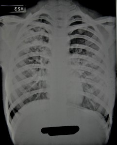
Paraplegia developed in September 1948 and he was transferred to the RVI that December showing signs of wasting and obvious kyphosis in the upper dorsal spine with paraplegia evident and total incontinence. His notes state that his head and thorax were encased in plaster cast, as was the left leg, to immobilise the affected areas. The incontinence was dealt with by applying a tube. Tuberculosis of the spine was relatively advanced, with the 3rd and 4th dorsal vertebrae having collapsed resulting in a noticeable kyphosis, seen in Figure 1.
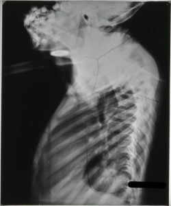
FIGURE 1: HOSP-STAN-07-01-02-1662-22

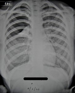
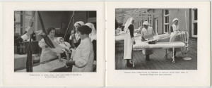
On admission to Stannington it is recorded that the radiographs showed a high dorsal lesion. The plaster cast encasing the head and thorax was removed and the patient was fixed to a short plaster boat with head piece, see left image in Figure 4.
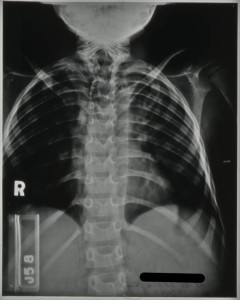
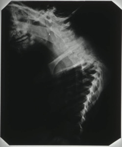
FIGURE 4: HOSP-STAN-07-01-02-1662-24
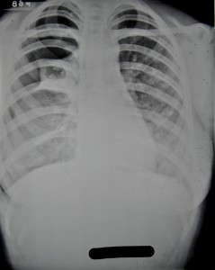
FIGURE 3: HOSP-STAN-07-01-02-1662-24
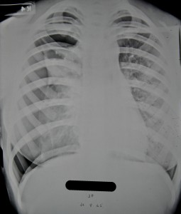
FIGURE 2: HOSP-STAN-07-01-02-1662-16


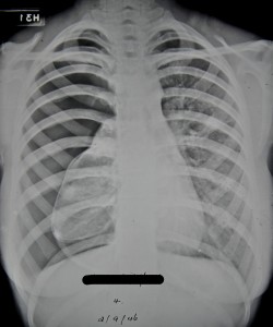
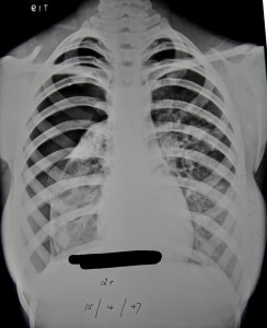
Radiographic images show further kyphosis, Figure 2, and the collapse of the vertebral bodies. Porosity is evident in the vertebral bodies in the upper dorsal region, identifiable by their translucent nature in Figure 3, giving rise to the extent of the infection.

In November 1949, the patient was fitted for a plastic splint. This was to fit

‘from the hips up the trunk extending over the neck to the occiput, reinforced with metal where necessary’.

The spinal lesion was considered quiescent by April 1952, all evidence of paraplegia having cleared up. However, he was to be fitted with a splint with a shaped head piece to immobilise the spine as much as possible. This patient was discharged in March 1953 and his brace discarded entirely in May 1953. He continued to be seen as an outpatient until February 1959.
HOSP-STAN-09-01-01
Figure 4:  HOSP-STAN-09-01-01


Case Study 2 – Cervical Vertebrae

Patient 148/1948, a 3 year old girl, was initially admitted in January 1948 (Patient Number 8/1948) with a Primary Tuberculous Complex of the right mid-zone.

Preliminary medical reports described this girl as having had an enlarged right hilar shadow, a shadow of the hilar lymph nodes, and ‘shotty,’ swollen, glands with an impetigious lesion on the scalp. However, her initial stay at Stannington was short as she was removed against medical advice by her mother 28 days after admittance, only to be re-admitted seven months later with TB of the cervical spine.

Following an examination by the surgeon, Mr Stanger, on re-admission a comprehensive outline of her condition was given:

The lower surface of the 2nd c.v (cervical vertebrae) is involved; the body of the 3rd c.v is completely destroyed and the upper surface of the 4th is probably eroded.

This child should have every bone in her body x-rayed.

The destruction of the vertebral bodies can be seen in the radiographs in Figures 5 and 6. Figure 5 shows the collapse of the vertebrae inwards creating a wedged shape in the neck. Figure 6, taken through the open mouth of the patient in order to gain a clear veiw of the vertebral bodies of the cervical vertebrae in the neck, shows a loss in denisty and clearly defined outer edges of the vertebral bodies due to collapse.
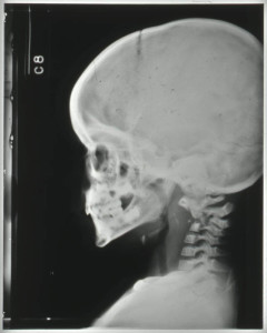
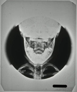
FIGURE 4: HOSP-STAN-07-01-02-1625-14
FIGURE 5: HOSP-STAN-07-01-02-1625-14
FIGURE 6: HOSP-STAN-07-01-02-1625-43


It is likely the request for all bones in her body to be x-rayed came from the suspicion that other areas of the skeleton had been affected by the disease. The request was carried out with the x-ray report card indicating that anteroposterior (AP) and lateral radiographs, where possible, were taken of the chest, spine, legs and hips. The patient was immobilised on a Bradford frame, a rectangular metal frame with canvas straps to hold the individual in a prone or supine position, seen in the right hand image of Figure 4.

Between September and December 1948 the patient is noted to have developed a number of additional symptoms, including vomiting sputum; patchy erythema (a scarlet rash) on her chest; purulent nasopharyngeal discharge (discharging pus from the nose); aural discharge; an inflamed throat and enlarged cervical glands.

By July 1949, these symptoms had largely been addressed and the patient was showing improvement. Immobilisation was considered satisfactory as a form of treatment and a moulded plastic splint was to be prepared for the patient, to consist of

a jacket taken from the hips and extending upwards to embrace the head and the occipital region to the chin.

This was later described as being reinforced with steel both vertically and transversely.


One year later, further examination by Mr Stanger noted that the disease had involved the 2nd, 3rd and 4th cervical vertebrae; the bodies of the 2nd and 3rd were showing signs of fusion and bone regeneration. It is at this point in July 1950, two years after first being admitted, that the child was allowed to ‘get up’.


This patient was discharged in December 1950, as being clinically and radiologically inactive and able to dispense with the splint. She continued to be seen as an outpatient at Stannington until 1956. Her last out-patient report stating that there was no deformity and no limitation of movement. Sound fusion was noted between both the vertebral bodies and posterior articulation of the 2nd, 3rd and 4th cervical vertebrae.


Further radiographic images can be seen on the Stannington Sanatorium ‘Radiographs from Stannington’ Flickr stream https://www.flickr.com/photos/99322319@N07/sets/72157648833066476/


Sources

Miller, F.J.W, Seal, R.M.E and Taylor, M.D (1963). Tuberculosis in Children. J & A Churchill Ltd.

Roberts, C and Buikstra, J (2003). The Bioarchaeology of Tuberculosis: A Global View on Reemerging Disease. Univesity Press of Florida.

Roberts, C and Manchester, K (2006). Archaeology of Disease (3rd Edition). Cornell University Press.

Manor Courts

The lord of the manor had the right to hold a court for his local tenants to facilitate management of the manor as a social and economic unit. From the beginning of the manorial system in the 11th century the manor courts conducted a variety of business. This was recorded on the court roll and by the 13th century it is evident that two main types of court are being recorded. The court baron, or ‘curia baronis’, was held every three weeks and handled the general business of the manor. This would involve issues relating to land tenure and use and enforce the payment of all dues and performance of services owed by the tenants to the lord. It also had other powers giving it jurisdiction over disputes between individuals and over personal actions by tenants, such as the recovery of small debts and complaints of trespass.

The court leet, or ‘curia leta’, was held every six months and inspected the working of the frankpledge, a system of mutual responsibility within a group of about ten households for the maintenance of law and order. This was often called the ‘view of frankpledge’. It also had powers to deal with offences such as common nuisances, affrays and the breaking of assize of bread and ale, (this regulated the price, weight and quantity of bread and beer sold). This court could fine and imprison offenders, in many manors in Northumberland the right went beyond imprisonment. The Barony of Langley was one of the lesser Baronies of Northumberland in which the Tindale family were enfeoffed, required to pledge service in exchange for land, by Henry I. They enjoyed an ancient liberty where they were able to try thieves in the Leet Court and then hang them on their own gallows.

The Barony of Embleton, via a succession of powerful Lords, had very extensive privileges. The incumbent Edmund Earl of Lancaster claimed, in 1292, the right to decide in his court pleas similar to those tried before the sheriff. He had a prison at Embleton and gallows at Newton, Embleton, Dunstan and Craster.

Woodhorn also seems to have had a licence for gallows in 1294, as well as Ovingham where in 1294 the Umfraville lordship claimed the right to pit and gallows, tumbrel, pillory and toll. Tynemouth also had the right to prison, gallows, tumbrel, and pillory and Bewick near Tynemouth a tumbrel and gallows.

Other types of court, which were held less frequently within the manor, included the court of survey and recognition, the court of pannage, the court of pie powder and the woodmote or forest court.

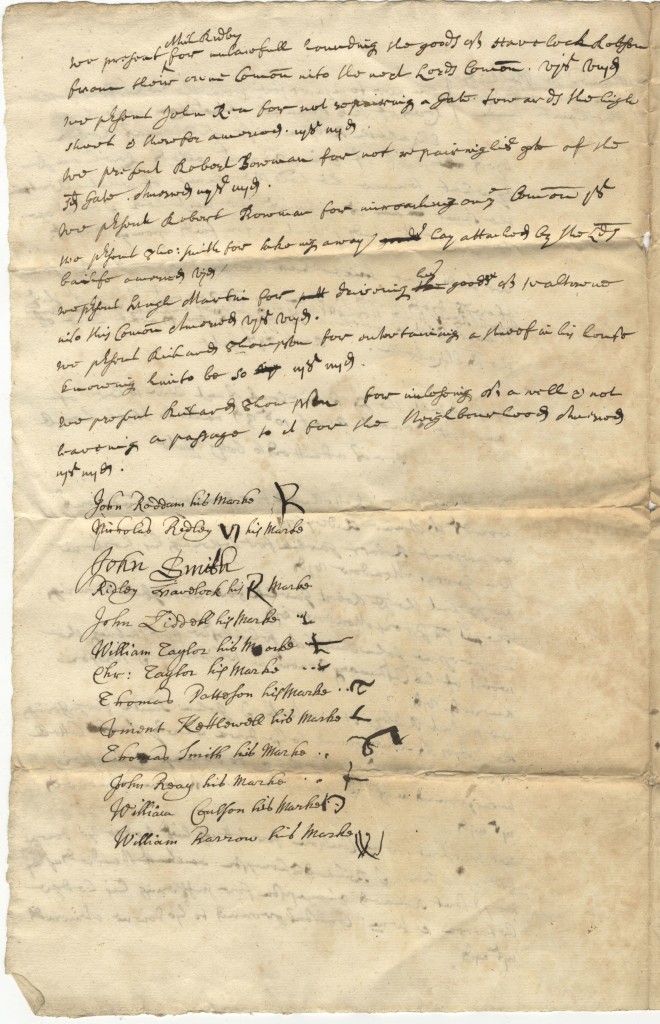
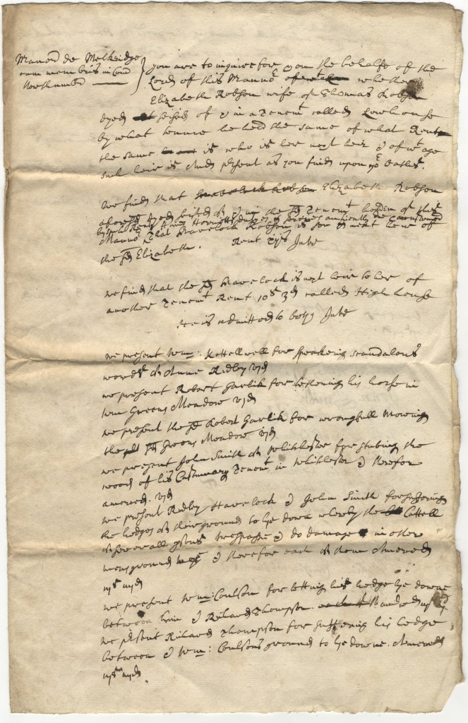
The business of the court was submitted via the presentments; this was done by the jury who were required to state or present the various matters which were dealt with by the court. The actual procedure for making presentments is not entirely clear and it is possible they were prepared several days in advance of the court session. The enclosed image is a presentment from the manor of Melkridge in Northumberland, dating from 1700, it gives a flavour of the type of court business being dealt with by the court baron.

ZBL 2/13/21
ZBL 2/13/21

[click to enlarge]

The jury are to enquire for and on behalf of the lord of the manor whether:

  • Elizabeth Robson wife of Thomas Robson died forfeit of and in a tenement called Lowhouse and to establish who and how old the heir is.
  • we present William Kettlewell for speaking scandalous words to Anne Ridley. vi d
  • we present Robert Garlick for tethering his horse in William Greens meadow. vi d
  • we present John Smith of Whitchester for steeling [?] the wood of his customary tenants in Whitchester and is therefore amerced (fined) vi d
  • we present Ridley Haverlock and John Smith for suffering the hedges of their ground to lie down whereby the cattle can damage other men’s grounds. iii s  iiii d


ZBL 2/13/21
ZBL 2/13/21

[click to enlarge]

  • we present John Rea for not repairing a gate towards the high shoot and is therefore amerced. iiis iiiid
  • we present Thomas Smith for taking away hay. vi d
  • we present Richard Thompson for entertaining a thief in his house and knowing him to be so. iiis iiiid
  • we present Richard Thompson for interfering with a well and not having a passage to it for the neighbourhood amerced. iiis iiiid

The presentment is signed by the jurors, note with the exception of John Smith, who can write his own name, most make their mark which in themselves are quite interesting as they have obviously been designed to be as unique as possible. A number of the jurors appear to be related to those on the presentment, or in the case of Ridley Havelock, seem to appear themselves.

Surgical Procedures – Artificial Pneumothorax

Pulmonary tuberculosis is by far the most common manifestation of TB witnessed throughout the Stannington records.  Prior to the development and use of any effective antibiotic treatments the most common form of intervention was the induction of an artificial pneumothorax.  Many of the different treatments employed to treat TB of all types at this time were based on the principles of resting and isolating the affected area, and the thinking behind artificial pneumothorax treatment was no different.


A needle would be inserted through the chest wall to allow for the insertion of air into the pleural cavity.  The amount of air inserted would depend on the size of the patient as well as how much the physician in charge though the patient could realistically manage in one go and how quickly they wished the lung to collapse.  Once inserted the pressure from the air would force the lung to collapse in on itself and to cease functioning properly.  The entire lung would not necessarily be collapsed at once, either because it wasn’t necessary for treatment or because fibrotic adhesions between the lung and the chest wall as a result of the disease prevented it from doing so.  Where only part of the lung was affected it would not be desirable to collapse the whole lung and in such instances just one lobe might be collapse.  Bilateral artificial pneumothorax was also a possibility, whereby part of both lungs would be collapsed at the same time.  A state of collapse could be maintained for a period of months or even years and required the patient to undergo regular refills of air in order to do so.


A great number of radiographic illustrations of the progression of a collapse are available in the Stannington collection.  One patient, 2/1946, has a large amount of radiographs taken over a period of two years which demonstrate the change in the lung from admission and through the progressive stages of lung collapse.


Patient 2/1946 was female an age15 when she was admitted to Stannington on 21 June 1945 with pulmonary TB stage 3, at which point her sputum tested positive for TB also.  A report on an x-ray taken pre-admission reads:

‘Right lung shows several active foci beginning to coalesce.  There is extensive infiltration in the upper zone & suspicious blotchy areas in the middle zone.  A small calcified opacity in the right lower zone.  The left lung shows infiltration in the middle zone.  The upper zone and apex are clear.  Early active foci are noticeable in both lungs in the affected areas.’

Figure 1 was the first x-ray taken after admission on 25 June 1945 being three weeks later than the one reported above.  Observations on this x-ray note:

‘Scattered foci in right upper zone.  One definite cavity.  Increased bronchial marking at both bases.’

HOSP/STAN/7/1/2/1057_22 25 June 1945
Figure 1 – HOSP/STAN/7/1/2/1057_22
25 June 1945
HOSP/STAN/7/1/2/1057_18 31 Aug 1945
Figure 2 – HOSP/STAN/7/1/2/1057_18
31 Aug 1945


It was quickly decided that and artificial pneumothorax should be induced on the right side and this took place on 16 Aug 1945. Figure 2 taken later on that month shows the initial results of the artificial pneumothorax.  The black area along the lateral side of the right lung is evidence of the air that has been inserted and the lung has begun to compress.


The collapse was maintained well into 1947 which involved her having refills of air every two weeks throughout this period.  For the first three months she received refills of 200-300ccs of air at a time, progressing to 400ccs the month after, and then eventually 500-600ccs at a time.  Figures 3-6 show the progression of the artificial pneumothorax as more air is inserted and the lung collapses further.  Over time we can see that the cavity in the right mid zone collapses and closes, one of the main aims of the treatment.  In early June 1946 a procedure was performed to divide adhesions between the lung and the chest wall which allowed the collapse to progress further.  She was discharged in June 1947 with her condition described as improved.


Figure 3 - HOSP/STAN/7/1/2/1057_23 17 Jan 1946
Figure 3 – HOSP/STAN/7/1/2/1057_23
17 Jan 1946
Figure 4 - HOSP/STAN/7/1/2/1057_09 18 June 1946
Figure 4 – HOSP/STAN/7/1/2/1057_09
18 June 1946


Figure 5 - HOSP/STAN/7/1/2/1057_10 2 Sept 1946
Figure 5 – HOSP/STAN/7/1/2/1057_10
2 Sept 1946
Figure 6 - HOSP/STAN/7/1/2/1057_27 15 April 1947
Figure 6 – HOSP/STAN/7/1/2/1057_27
15 April 1947